Non-damage micrometering system for observating biological sample with fluorescent

A biological sample, non-destructive technology, applied in the field of electrochemistry, can solve the problem of inability to observe the fluorescence of biological samples

Inactive Publication Date: 2009-02-11
CAPITAL NORMAL UNIVERSITY
View PDF0 Cites 19 Cited by
  • Summary
  • Abstract
  • Description
  • Claims
  • Application Information

AI Technical Summary

Problems solved by technology

[0011] In order to overcome the disadvantage that the optical imaging system of the existing non-destructive micro-measurement system cannot observe the fluorescence emitted by biological samples, the present invention improves the optical imaging system on the basis of the existing non-destructive micro-measureme

Method used


Image

  • Non-damage micrometering system for observating biological sample with fluorescent
  • Non-damage micrometering system for observating biological sample with fluorescent
  • Non-damage micrometering system for observating biological sample with fluorescent

Examples


example 1

[0028] Example 1: A non-invasive micro-measurement system capable of observing and sorting fluorescent biological samples

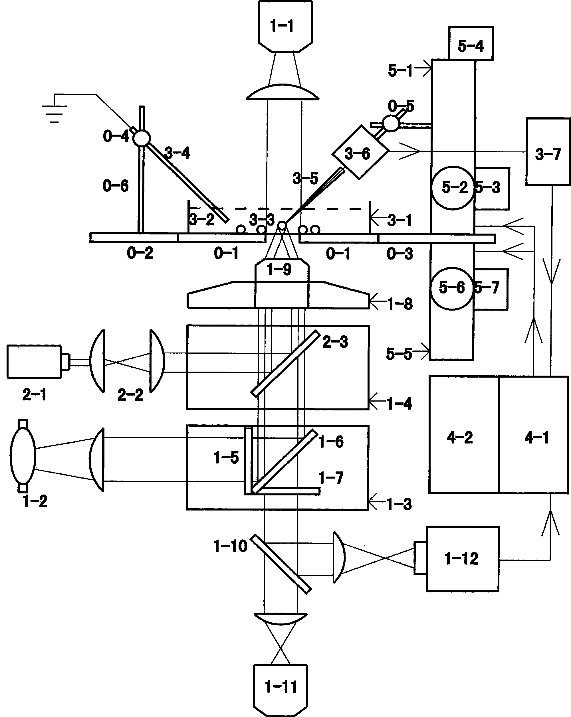
[0029] figure 1 The illustrated embodiment is a non-invasive micro-measurement system that combines a fluorescent device and an optical clamp device, which can be used for fluorescent identification of transformed biological samples with green fluorescent protein (GFP) markers and optical clamp fixation and movement of yeast cells.

[0030] Among them, bright field illuminator 1-1, fluorescent illuminator 1-2, stage 0-1, objective lens converter 1-8, filter turret converter 1-3, filter turret converter 1-4, Structures such as eyepieces 1-11 are provided by commercial manufacturers of inverted fluorescence microscopes. A 100-fold objective lens is installed in the objective lens converter 1-8 to converge the clamp laser beam. The filter turntable converter 1-3 is equipped with: excitation filter 1-5 has a wavelength of 450-490 nanometers, and limits the exci...

Embodiment 2

[0035] Example 2: Application of the present invention in the detection of genetically modified plant materials

[0036] 1. Transgenic plant material

[0037] GFP fusion protein transgenic Arabidopsis thaliana seedlings germinated for 5 days

[0038] 2. Screening and image capture of materials by fluorescence microscope

[0039] The roots, root tips, and root hairs of the transgenic plants were clearly observed under a 10x or 40x microscope in the bright field. Then it is converted to blue excitation light to excite the sample, and the transgenic material with green fluorescent expression is observed and captured. Finally, use the software to capture the image.

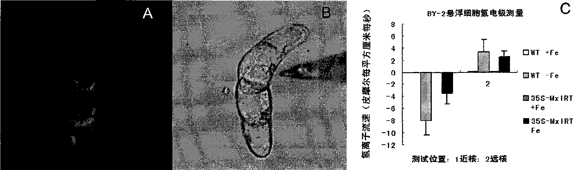
[0040] 3. Non-damaging micro-test

[0041] Including: glass electrode drawing, molecular / ion electrode buffer filling, LIX filling, ion / molecular electrode silver wire chlorination, molecular / ion electrode static calibration, and testing of experimental samples. The part tested in the experiment: the root hair area (such ...

Embodiment 3

[0044] Example 3: The present invention is used for the detection of transgenic tobacco suspension cells

[0045] 1. Cultivation and fixation of transgenic tobacco suspension cells

[0046] Transgenic tobacco suspension cells that have been screened for resistance are subcultured with sterile modified MS liquid medium at a ratio of 1:10 once a week, and can be used for detection 3-4 days after subculture. Improved MS medium is newly added with KH 2 PO 4 30mg / L, 2,4-D 0.6mg / L, VB1 0.5mg / L MS medium. During the test, 1% low melting point glue was used to fix the transgenic tobacco suspension cells in the test container.

[0047] 2. Screening and image capture of materials by fluorescence microscope

[0048] In bright field, clear transgenic tobacco suspension cells were observed with a 10x lens or a 40x lens. Then the sample was excited by blue excitation light, and the transgenic tobacco suspension cells expressing green fluorescence were observed as the capture object. Finally, us...


PUM


Abstract

In order to overcome the shortcomings of the optical imaging system of available non-destructive micro-measuring system on the inability to observe the fluorescence emitted from a biological sample, The invention modifies the optical imaging system based on the available non-destructive micro-measuring system so that the non-destructive micro-measuring system can observe the fluorescence emitted by the biological sample while measuring the ion/molecule flowing direction on the surface of a biomaterial to implement the screening and the state monitoring of the biomaterial to be measured. The invention belongs to the electrochemical technical field, and comprises an ion/molecule activity detection system, a micro-imaging system and a computer data processing system. The micro-imaging system includes a bright field illuminator, a stage, an objective converter, a light path switcher, an eyepiece and a digital camera, and is characterized in that an optical filter turntable converter for a fluorescence microscopic observation mode is disposed on the light path between the objective and the light path switcher, and includes an excitation optical filter, a fluorescent dichroic mirror and a blocking filter.

Description

Technical field [0001] The invention belongs to the field of electrochemistry technology, and specifically relates to an improved optical electrophysiology system capable of detecting the flow rate and direction of ion / molecule transport. The application of the invention can observe the electrophysiological condition of cells. Background technique [0002] Substances have a tendency to diffuse from high concentration to low concentration in the liquid environment. For charged particles, there is also a tendency to move from a high electrochemical potential to a low electrochemical potential. [0003] The core of the non-damaging micro-measurement system is the selective ion / molecular microelectrode, hereinafter referred to as the ion / molecule selective electrode, which is developed by a computer-controlled automatic positioning measurement system designed by Kühtreiber and Jaffe in 1990 (Kühtreiber And Jaffe in J. Cell Biol 1990, 110: 1565-1573). The ion selective electrode is us...

Claims


Application Information

Patent Timeline
IPC IPC(8): G01N33/48G01N21/64C12Q1/02
Inventor 印莉萍赵伟忠杨光郭家峰靳思房茜张鹏
Owner CAPITAL NORMAL UNIVERSITY
Why Eureka
Try Eureka