
Nanoparticle probes for detecting ribosome inactivating protein, manufacturing method thereof and use thereof

A ribosome inactivation and nanoparticle technology is applied in the field of nanoparticle probes and their preparation, which can solve the problems of complicated steps and low sensitivity, and achieve the effects of simple development process, high sensitivity, and easy popularization and application.

Inactive Publication Date: 2010-06-16
NAT INST OF METROLOGY CHINA
View PDF0 Cites 4 Cited by
  • Summary
  • Abstract
  • Description
  • Claims
  • Application Information

AI Technical Summary

Problems solved by technology

[0010] The primary problem to be solved by the present invention is the low sensitivity and cumbersome steps of the traditional ribosome-inactivating protein detection method

Method used


Image

  • Nanoparticle probes for detecting ribosome inactivating protein, manufacturing method thereof and use thereof
  • Nanoparticle probes for detecting ribosome inactivating protein, manufacturing method thereof and use thereof
  • Nanoparticle probes for detecting ribosome inactivating protein, manufacturing method thereof and use thereof

Examples


preparation example Construction

[0057] ③ Preparation of nanoparticle probes

[0058] The antibody modified by the binding site is reacted with the surface-modified matrix nanoparticles at a constant temperature, the reaction temperature is 20-50°C, the reaction time is 0.5-5h, and the pH value is between 7.0-8.5, and the specific antibody is obtained. nanoparticle probes.

[0059] ④ Separation of nanoparticle probes

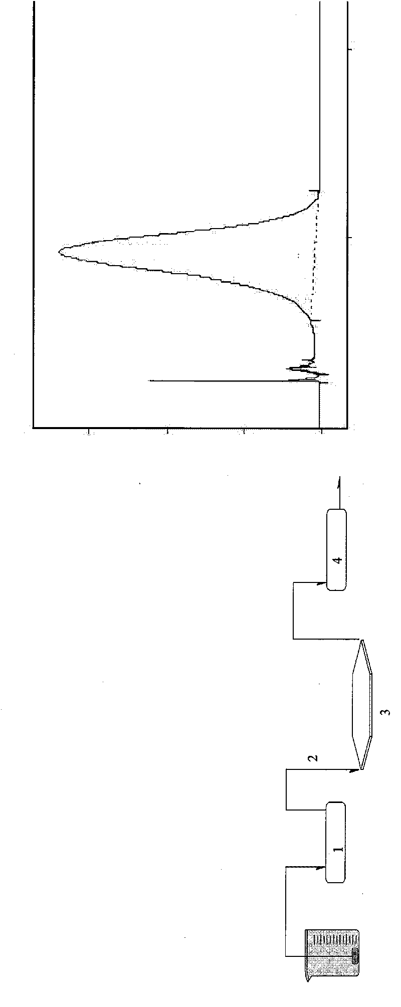
[0060] Using sedimentation field flow separation technology, it can separate suspended particles in the range of 20nm-1μm in the fluid. Using the mobile phase containing FL-70 surfactant, under the action of external fields such as centrifugal force field, according to the difference in particle size and quality, Nanoparticles of different particle sizes are distributed in a certain way in the thickness of the channel, and the fluid near the channel wall will flow slower than in the center; the difference in the flow velocity of nanoparticles of different particle sizes in the channel will lea...

Embodiment 1

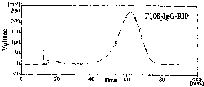
[0085] Nanoparticle probe and its preparation for detection of ricin

[0086] ① Surface modification of polystyrene matrix nanoparticles by modifier F108-PDS

[0087] Proportionally weigh a certain volume of modifier F108-PDS (0.01-10g / L) and add it to the polystyrene nanoparticle suspension with a particle size of 290nm. Modifiers, and then centrifuge under certain conditions to separate the modifiers that are not bound or not tightly bound, discard the supernatant and re-dilute the nanoparticles with phosphate buffer.

[0088] ②Chemical modification of mouse anti-human IgG monoclonal antibody sites

[0089] Use n-hydroxysuccinimide 3-(2-pyridyldimercapto) propionate (concentration: 0.01-5g / L) to modify the active site of the antibody, and weigh them respectively according to the ratio of the two (0.1-1.5). Take a certain volume (1-20 μL) of the modifier and add it to the antibody solution. After reacting at a temperature of 20-50°C, use a specific chromatographic column t...


PUM

PropertyMeasurementUnit
Particle sizeaaaaaaaaaa

Abstract

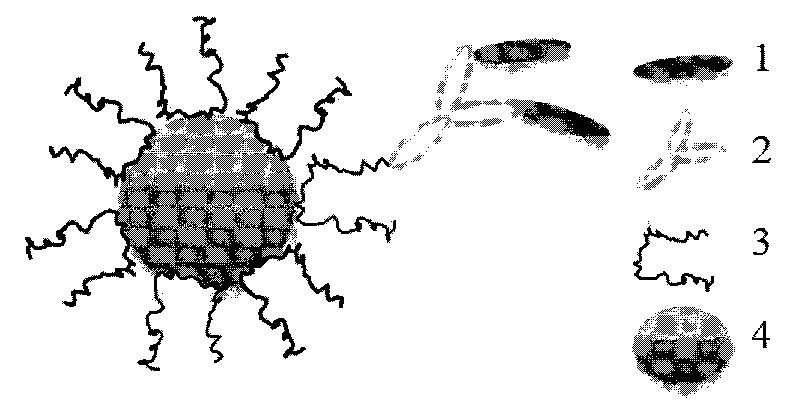
The invention provides nanoparticle probes for detecting ribosome inactivating protein, a manufacturing method thereof and use thereof. In the nanoparticle probes, nanoparticles with bioaffinity serving as a matrix can be combined with a target protein antibody after undergoing surface modification, and the interference of physical absorption is eliminated; and a monoclonal antibody of rat anti-human immunoglobulin IgG serving as a capture antibody is used to be coupled with a target ribosome inactivating protein through the specific interacting force between the antibody and the antigen. Thenanoparticle probes for detecting the ribosome inactivating protein of the invention combine the high concentrating capability of the nanoparticles with large specific surface area with the high antigen selectivity of the monoclonal antibody, so the sensitivity and specificity of the detection of the ribosome inactivating protein are improved. The nanoparticle probes of the invention comprehensively use nano technology and immunity technology and are applied to the detection of the target ribosome inactivating protein in the fields of food safety, anti-bioterrorism and the like. In addition, the nanoparticle probes have the advantages of quickness, convenient carrying, specific trace detection and the like.

Description

technical field [0001] The invention relates to a nanoparticle probe used for detecting ribosome inactivation protein and a preparation method thereof, and belongs to the fields of nanotechnology, food safety, biotechnology application and the like. Background technique [0002] Ribosome-inactivating protein (Ribosome-inactivating protein, RIP) exists in many plants, and the physiological function of plant ribosome-inactivating protein is to play a defensive role, that is, to resist diseases and insect pests or harsh environments. According to the primary structure of ribosome-inactivating protein, RIP can be divided into the following two types: type I, consisting of one polypeptide chain; type II, two-chain protein, consisting of two peptide chains. In the two-chain protein, one of them is the A (active) chain, which has N-glycosidase activity; the other is the B (binding) chain, which is a lectin specific for galactose, and the two polypeptide chains pass disulfide bonds ...

Claims


Application Information

Patent Timeline
IPC IPC(8): G01N33/68G01N33/577G01N33/553G01N33/531
Inventor 全灿刘军李红梅
Owner NAT INST OF METROLOGY CHINA
Learn More