Novel in situ liquid crystal vascular embolization agent

A vascular embolizing agent and in-situ liquid crystal technology, which is applied in the field of in-situ liquid crystal vascular embolic agents and vascular embolizing agents for interventional therapy, can solve the problems of difficulty in preparation, strict requirements on phase transition temperature, etc.

Inactive Publication Date: 2010-09-08
广州万泽医药科技有限公司
View PDF6 Cites 24 Cited by
  • Summary
  • Abstract
  • Description
  • Claims
  • Application Information

AI Technical Summary

Problems solved by technology

Therefore, the injectable temperature-sensitive hydrogel material has attracted the attention of researchers, and its response to temperature makes it an ideal drug delivery carrier and tissue engineering scaffold material. However, because it is a temperature-sensitive material, Therefore, the phase transition temperature is strictly required, which brings difficulties to the preparation, and the microcatheter needs to be cooled during embolization to prevent the phase transition of the material in the microtube, which limits its application.

Method used


Image

  • Novel in situ liquid crystal vascular embolization agent
  • Novel in situ liquid crystal vascular embolization agent
  • Novel in situ liquid crystal vascular embolization agent

Examples


Embodiment 1

[0018] The preparation of the in-situ liquid crystal vascular embolism described in this example is as follows: Weigh 1.2 g of absolute ethanol and 3.2 g of phytantriol, vortex and mix both, add 0.5 g of water, vortex mix, and centrifuge to obtain a clear Liquid embolic agents.

[0019] New Zealand rabbits were anesthetized by ear vein injection with 0.2ml / kg Sumianxin. After the rabbits were anesthetized for 5 to 10 minutes, they were placed on the self-made fixing board, and the board was placed on the DSA operating table. The right groin Hair removal and disinfection, sterile surgical drape, incision of the groin skin, separation to the femoral arteriovenous sheath layer by layer, intermittent spraying of lidocaine, exposure of the right femoral artery and hemostatic forceps to separate and expose the femoral artery, both ends covered with silk thread , Lift both ends of the silk thread to separate the femoral artery from the surrounding tissue. Then puncture the femoral a...

Embodiment 2

[0021] The preparation of the in-situ liquid crystal vascular embolization agent described in this example is as follows: Weigh 1.2 g of absolute ethanol and 3.2 g of phytantriol, vortex and mix both, add 1 g of water, vortex mix, and centrifuge to obtain a clarified Liquid embolic agents.

[0022] The obtained liquid embolic agent is vortex-mixed with 1 g of lipiodolized oil to obtain a contrastable liquid embolic agent. The resulting contrastable liquid embolic agent is used to embolize the hepatic arteries of rabbits (method is the same as in Example 1), as figure 2 As shown, it has developing and embolic effects.

Embodiment 3

[0024] Establish VX2 liver cancer tumor-bearing rabbits.

[0025] The preparation of the in-situ liquid crystal vascular embolization agent described in this example is as follows: Weigh 1.8 g of absolute ethanol, add 5 mg of docetaxel, and then add 2.7 g of glycerol monooleate, vortex and mix the two (slightly heated), Add 0.5g of water, vortex mix (slightly heated), and centrifuge to obtain a clear liquid embolism.

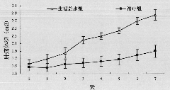
[0026] After the liquid embolic agent of gained is added 3g iodized oil, rabbit is carried out hepatic artery embolization (method is the same as embodiment 1), observes tumor volume change under CT, see image 3 (with the normal saline group as the control), the results show that the in situ liquid crystal blood vessel embolization agent can effectively inhibit tumor growth. In addition, there was no tube blockage during the experiment, and the animals moved normally.


PUM


Abstract

The invention discloses a novel in situ liquid crystal vascular embolization agent, which mainly comprises a liquid crystal material, a water-soluble physiological acceptable solvent and water, wherein the ratio of mass parts of the liquid crystal material, the water-soluble solvent and the water is 0.3-3.2:1.2-2.1:0.5-1, and the mass percent of the water in the vascular embolization agent is less than 50%; and the liquid crystal material is of an amphiphilic polymer. The in situ liquid crystal vascular embolization agent has the effects of non-toxicity, non-adhesion, easy operation, safety, effectiveness and low cost, and the characteristics of good in vitro fluidity and fast sol-gel conversion speed.

Description

technical field [0001] The invention relates to the field of cancer palliative treatment, in particular to a new type of vascular embolism for interventional therapy—in-situ liquid crystal vascular embolism. Background technique [0002] Cancer is a common disease that seriously endangers human life and health. According to the report of the World Health Organization, the incidence and mortality of cancer in the world are increasing year by year. In my country, cancer has become the second leading cause of death after cardiovascular disease, and its late discovery and difficult cure have become important causes of death from this disease. Therefore, the research on early diagnosis and treatment of cancer has always been a hot spot, especially in diagnosis and therapeutics, which has aroused extensive interest of researchers. [0003] The treatment of cancer is a systematic project, which cannot be satisfied with only specialized local treatment methods, and the era of surge...

Claims


Application Information

Patent Timeline
Patent Type & Authority Applications(China)
IPC IPC(8): A61K9/00A61K47/30A61K47/14A61K47/10A61K47/22A61K47/34A61K45/00A61K33/24A61K31/337A61P35/00
Inventor 吴传斌韩珂
Owner 广州万泽医药科技有限公司
Why Eureka
Try Eureka