Tissue engineered bone cartilage frame and preparation method thereof

A tissue engineering bone and osteochondral technology, applied in the field of tissue engineering osteochondral scaffolds and constructing osteochondral complexes, can solve problems such as interface mechanical defects, poor mechanical properties of upper materials, poor shear resistance, etc., and achieve good flexibility and mechanical properties, reducing antigenicity, and beneficial to osteogenesis

Inactive Publication Date: 2010-09-08
中国人民解放军第三0九医院
View PDF8 Cites 10 Cited by
  • Summary
  • Abstract
  • Description
  • Claims
  • Application Information

AI Technical Summary

Problems solved by technology

(2) The fixation problem of tissue engineered articular cartilage products has not been effectively solved
(3) Existing tissue engineered cartilage has biomechanical defects
(4) Poor integration between tissue engineered cartilage and subchondral bone
[0005] The composite osteochondral scaffold using the natural gel-like material as the upper cartilage scaffold has the following problems: ①The upper layer material has poor mechanical properties; ②There are mechanical defects between the interfaces, which are easy to tear off and have poor shear resistance; ③Gel-like scaffolds After implanting in the body, it absorbs water and deforms, turning into a gel again, and the mechanical properties of the cartilage layer are further weakened; ④The operation is relatively complicated; ⑤The lower layer of hydroxyapatite degrades slowly, which affects the healing with the surrounding bone
[0010] There are many studies and patents on the application of decalcified bone matrix as a scaffold for cartilage tissue engineering, but they are all only used for cartilage tissue engineering, and the aforementioned fixation problems still exist when implanted in the body

Method used


Image

  • Tissue engineered bone cartilage frame and preparation method thereof
  • Tissue engineered bone cartilage frame and preparation method thereof
  • Tissue engineered bone cartilage frame and preparation method thereof

Examples


Embodiment 1

[0035] Embodiment 1 uses fresh bovine femoral condyle as raw material to prepare tissue engineered osteochondral scaffold

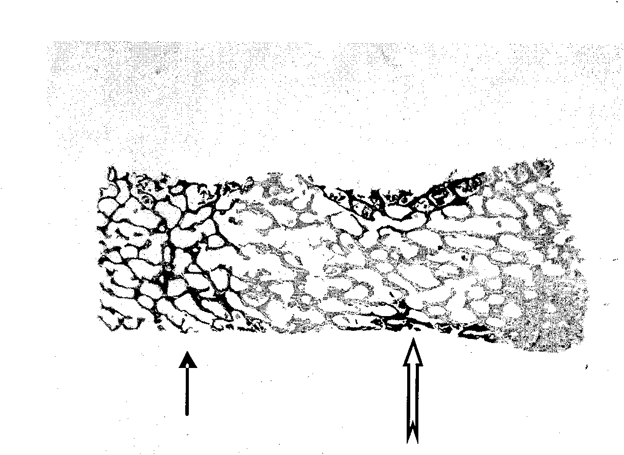
[0036] Purchasing fresh bovine femoral condyles from the market, removing the attached muscles and ligaments, sawing them into 2cm thick bone slices, washing the bone marrow with a high-pressure water gun and triple distilled water, cutting off the bone cortex, and dehydrating with absolute ethanol , degreased with ether, and air-dried. Then, a 4mm diameter trepan was used to make a cancellous bone column with a diameter of 4mm from the treated bovine femoral condyle. The lower 2 / 3 of the cancellous bone column was embedded in paraffin. Place the cancellous bone column as a whole in 10% (w / v) EDTA solution for decalcification for 2 weeks, then remove paraffin with xylene and graded alcohol, soak in PBS buffer solution for more than 1 week, and then use a freeze dryer according to the instructions By freeze-drying, the layered decalcified tissue engineer...

Embodiment 2

[0037] Example 2 Using Fresh Bovine Femoral Condyle as Raw Material to Prepare Tissue Engineering Osteochondral Scaffold

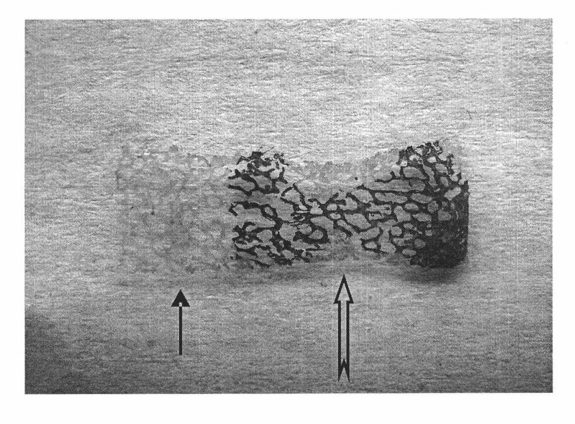
[0038] Purchasing fresh bovine femoral condyles from the market, removing the attached muscles and ligaments, sawing them into 2cm thick bone slices, washing the bone marrow with a high-pressure water gun and triple distilled water, cutting off the bone cortex, and dehydrating with absolute ethanol , degreased with ether, and air-dried. Then a trephine with a diameter of 1 cm was used to make a cancellous bone column with a diameter of 1 cm from the treated bovine femoral condyle. The lower 2 / 3 of the cancellous bone column was embedded in paraffin. The lower 2 / 3 part of the paraffin-embedded cancellous bone column was placed in 10% (w / v) EDTA solution for decalcification for 2 weeks, then the paraffin was removed with xylene and graded alcohol, and soaked in PBS buffer for more than 1 week. Then freeze-dried,.

Embodiment 3

[0039] Example 3 Using Fresh Bovine Femoral Condyle as Raw Material to Prepare Tissue Engineering Osteochondral Scaffold

[0040] Purchasing fresh bovine femoral condyles from the market, removing the attached muscles and ligaments, sawing them into 2cm thick bone slices, washing the bone marrow with a high-pressure water gun and triple distilled water, cutting off the bone cortex, and dehydrating with absolute ethanol , degreased with ether, and air-dried. Then, a 2 cm diameter trepan was used to make a cancellous bone column with a diameter of 2 cm from the treated bovine femoral condyle. The lower 2 / 3 of the cancellous bone column was embedded in paraffin. The lower 2 / 3 part of the paraffin-embedded cancellous bone column was placed in 10% (w / v) EDTA solution for decalcification for 2 weeks, then the paraffin was removed with xylene and graded alcohol, and soaked in PBS buffer for more than 1 week. Then freeze dry.


PUM

PropertyMeasurementUnit
diameteraaaaaaaaaa
diameteraaaaaaaaaa
diameteraaaaaaaaaa

Abstract

The invention belongs to the field of cartilage tissue repair and tissue engineering and provides a tissue engineered bone cartilage frame and a preparation method thereof. Particularly, marrow removal and degreasing are carried out on allogeneic or xenogeneic cancellous bones to obtain cancellous bone columns with experimentally or clinically required diameters, and then a tissue engineered bonecartilage frame with a completely decalcified upper layer and a superficially decalcified lower layer is obtained via layered decalcification. The upper layer of the frame is used as the framework part of the tissue engineered cartilage frame or an engineering cartilage frame, and the lower layer thereof can be used as a bone forming frame, an engineering bone bracket and the fixed pivot of the engineered cartilage, and can be used for constructing an integrative tissue engineered bone cartilage or directly transplanted for the cartilage or bone cartilage repair. Compared with the existing tissue engineered bone cartilage frame, the invention has the advantages of integration (no interface mechanical defect), and good compatibility of a pore structure and the tissue, has better compression resisting, tensile resisting and shear resisting properties, and is convenient for the minimally invasive surgery operation under an arthroscope.

Description

technical field [0001] The invention belongs to the field of cartilage tissue repair and tissue engineering. The invention provides a method for preparing tissue engineered osteochondral scaffold and the tissue engineered osteochondral scaffold prepared by the method. Specifically, the present invention uses cancellous bone as a raw material and utilizes layered decalcification technology to prepare tissue engineered osteochondral scaffolds, and the prepared tissue engineered osteochondral scaffolds can be used to construct osteochondral complexes. Background technique [0002] Injuries and lesions of articular cartilage are common diseases, and it is difficult to repair by itself. Tissue-engineered cartilage is the most promising repair method. However, the following problems need to be solved urgently in the research of tissue engineering of articular cartilage: (1) When repairing articular cartilage, it is often necessary to repair subchondral bone. Clinically, simple ...

Claims


Application Information

Patent Timeline
Patent Type & Authority Applications(China)
IPC IPC(8): A61L27/36
Inventor 余方圆黄利虹马远征刘海容
Owner 中国人民解放军第三0九医院
Why Eureka
Try Eureka