Specific detection method of protein or polypeptide cysteine sulfydryl modification and application thereof

A technology of cysteine ​​sulfhydryl and cystine sulfhydryl, which is applied in the field of detection of post-translational modification of proteins or polypeptides, and can solve problems such as interference

Active Publication Date: 2010-11-24
INSITUTE OF BIOPHYSICS CHINESE ACADEMY OF SCIENCES
View PDF8 Cites 31 Cited by
  • Summary
  • Abstract
  • Description
  • Claims
  • Application Information

AI Technical Summary

Problems solved by technology

This suggests that the method is not specific for the detection of protein thiopalmitoylation: intermolecular disulfide bonds interfere with the detection of protein thiopalmitoylation by this method

Method used


Image

  • Specific detection method of protein or polypeptide cysteine sulfydryl modification and application thereof
  • Specific detection method of protein or polypeptide cysteine sulfydryl modification and application thereof
  • Specific detection method of protein or polypeptide cysteine sulfydryl modification and application thereof

Examples


Embodiment 1

[0106] Example 1: Materials and methods

[0107] animal

[0108] We used 4-5 week old C57BL / 6 mice (SPF grade, Beijing Weitong Lihua Experimental Animal Technology Co., Ltd.). All animal-related procedures were approved by the Animal Care Committee of the Institute of Biophysics, Chinese Academy of Sciences.

[0109] Raw Biotin Conversion Method

[0110]Mouse brain was homogenized using 5ml of HEN buffer (25mM HEPES (4-(2-hydroxyerhyl)piperazine-1-erhanesulfonic acid, 4-hydroxyethylpiperazineethanesulfonic acid) pH 7.7, 0.1mM EDTA (ethylenediamine Tetraacetic acid), 10mM neocuproine (2,9-dimethyl-1,10-phenanthroline)), add 1% NP40 (Nonidet P-40, ethylphenyl polyethylene glycol) in the homogenate, protease inhibition reagent, 1 mM PMSF (phenylmethylsulfonyl fluoride), centrifuged at 12,000 g at 4°C for 10 minutes. Half of the supernatant was treated with 40 μM NO donor GSNO (S-nitrosoglutathione) for 30 minutes at room temperature. The NO donor was removed by acetone preci...

Embodiment 2

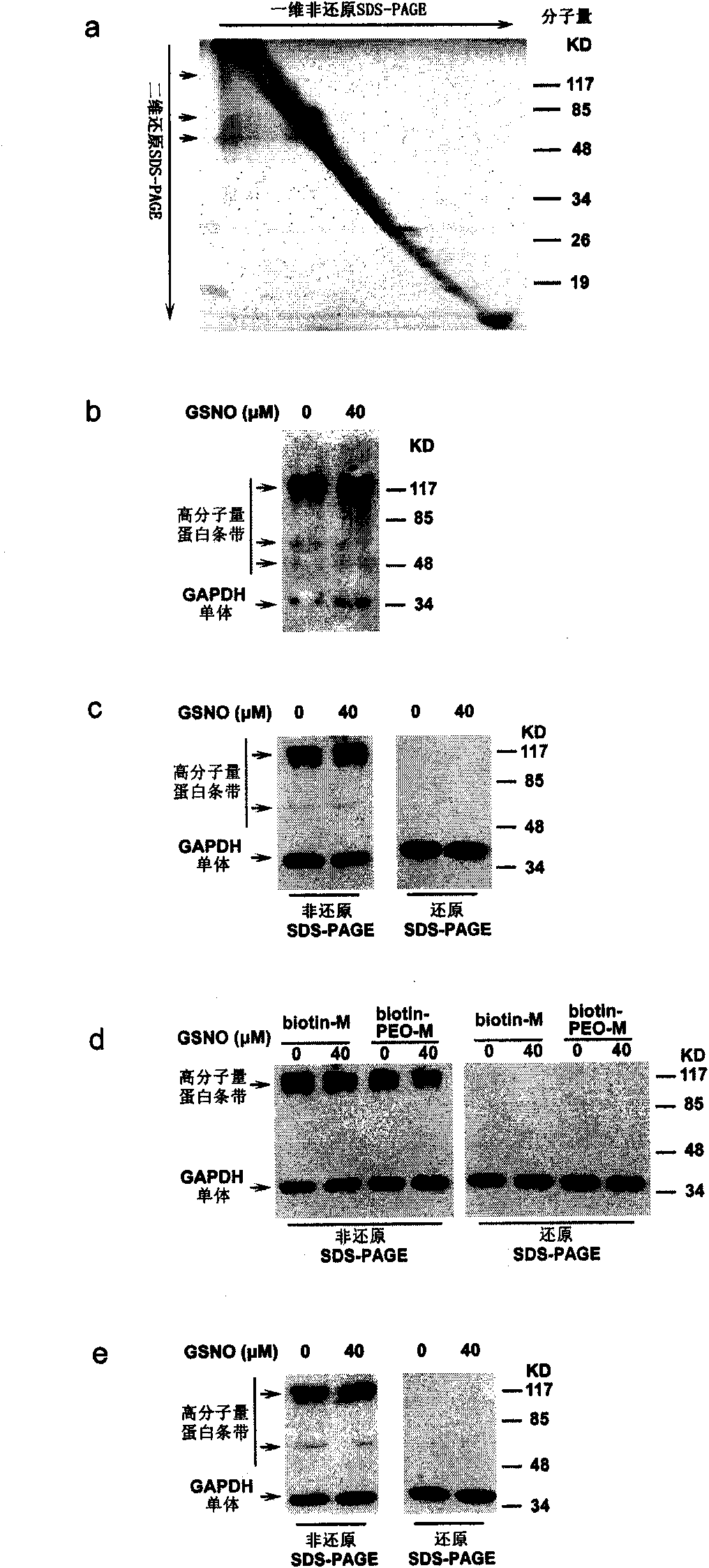
[0117] Example 2 uses "diagonal eleetrophoresis" 10,11 and Western blot analysis to detect the original biotin conversion method is interfered by intermolecular disulfide bonds ( image 3 )

[0118] After the first-dimension non-reducing SDS-PAGE, the entire electrophoresis lane was excised for the second-dimension reducing SDS-PAGE. If there is no intermolecular disulfide bond, the molecular weight of the protein band will not change, and it will be on a diagonal; if there is an intermolecular disulfide bond, then the molecular weight of the protein will decrease and move below the diagonal 10,11 . When eluting protein samples that have undergone the biotin conversion method, we use a non-reducing denaturing elution method: high temperature and high concentration of denaturant. This ensures that after elution, potential intermolecular disulfide bonds remain intact and can be analyzed by subsequent diagonal electrophoresis.

[0119] We treated mouse brain homogenates with...

Embodiment 3

[0121] Example 3 uses different sulfhydryl blocking reagents, purification reagents, and biotinylation reagents. Intermolecular disulfide bonds exist in all cases ( Figure 4 )

[0122] We doubted whether these intermolecular disulfide bonds were formed during the MMTS-blocking step, so we replaced the reversible thiol-blocking reagent MMTS with an irreversible thiol-blocking reagent, NEM (N-ehylmaleimide), but we Intermolecular disulfide bonds were still found in subsequent protein samples ( Figure 4 a). Then we replaced the biotinylation reagent biotin-HPDP with biotin-maleimide or the purification reagent streptavidin sepharose beads with avidin sepharose beads, the intermolecular disulfide bond still exists ( Figure 4 b, Figure 4 c). These results imply that the detected intermolecular disulfide bonds are not derived from the biotin conversion process, but from the original sample of mouse brain homogenate.


PUM


Abstract

The invention relates to a specific detection method of protein or polypeptide cysteine sulfydryl related modification under the condition that the interference of the intermolecular disulfide bond is removed, a kit for executing the method and application of the kit in screening new physiopathology related endogenous targets or exogenous protein or polypeptide targets of cysteine sulfydryl related modification, screening drugs designed to regulate the protein or polypeptide cysteine sulfydryl related modification and detecting content of cysteine sulfydryl related modification in samples.

Description

technical field [0001] The present invention relates to the detection field of protein or polypeptide post-translational modification, in particular, to the detection field of protein or polypeptide cysteine ​​sulfhydryl-related modification, more specifically, to a method for detecting protein or polypeptide cysteine ​​sulfhydryl-related modification and Use thereof, and kits for performing the method. Background technique [0002] Cysteine ​​(cys) on a protein or polypeptide contains a free sulfhydryl group (-SH). This sulfhydryl group can undergo various protein post-translational modifications. Each modification has a specific mode of regulation and biological role. With the development of proteomics, methods for high-throughput screening of protein targets with these modifications have been gradually developed. In fact, several of these modifications can be detected using the same principle. Their principles are all based on the method of "biotin switch" followed by...

Claims


Application Information

Patent Timeline
Patent Type & Authority Applications(China)
IPC IPC(8): G01N33/68G01N27/447
Inventor 陈畅黄波
Owner INSITUTE OF BIOPHYSICS CHINESE ACADEMY OF SCIENCES
Why Eureka
Try Eureka