Inhibitor for inhibiting expression of MBD2 and application thereof

An inhibitor and molecular technology, applied in the field of molecular biology and biomedicine, which can solve the problems of different affinities, embryo death, and inability to selectively recognize methylated DNA.

Inactive Publication Date: 2010-12-15
TONGJI HOSPITAL ATTACHED TO TONGJI MEDICAL COLLEGE HUAZHONG SCI TECH
View PDF3 Cites 3 Cited by
  • Summary
  • Abstract
  • Description
  • Claims
  • Application Information

AI Technical Summary

Problems solved by technology

The affinity of each MBD protein to methylated DNA is different, and the highest affinity is the MBD2 protein, but MBD3 in mammals cannot selectively re

Method used


Image

  • Inhibitor for inhibiting expression of MBD2 and application thereof
  • Inhibitor for inhibiting expression of MBD2 and application thereof
  • Inhibitor for inhibiting expression of MBD2 and application thereof

Examples


Embodiment 1

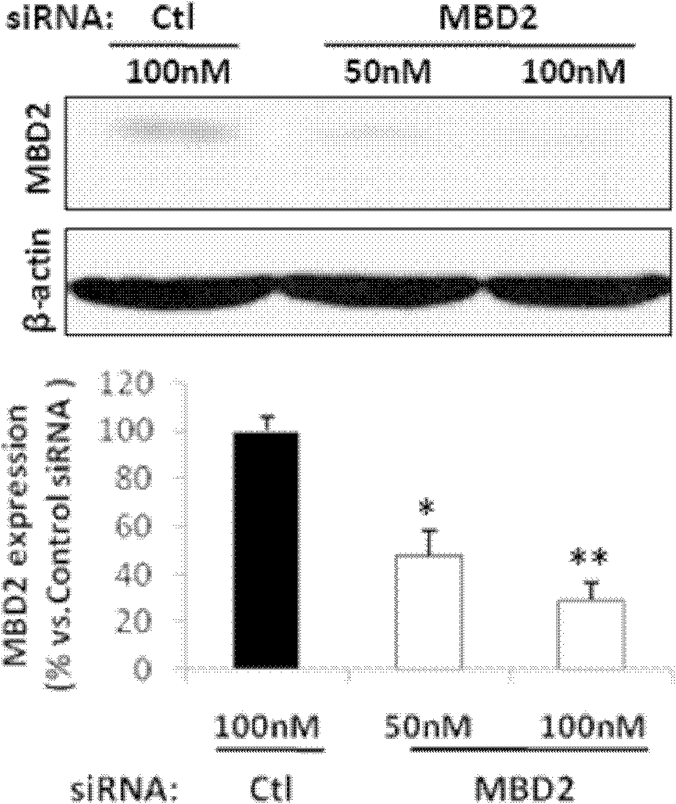
[0038] Example 1: MBD2 siRNA inhibits the expression of MBD2 protein in human umbilical vein endothelial cells

[0039] Human Umbilical Vein Endothelial Cells (HUVECs) were cultured in EBM-2 epithelial cell medium in 5% CO 2 , Cultured in a 37°C incubator. Cells were seeded in 12-well culture plates 24 hours before transfection at a seeding density of 4×10 4 , the next day, transfection was performed according to the Lipofectamine (Invitrogen, Carlsbad, CA) reagent operating instructions, and 50, 100 nmol / LMBD2 siRNA and a control group were set. Cells were collected 24-48 hours after transfection, and proteins were extracted for immunoblotting. Proteins were separated on 8% SDS-PAGE gel and transferred to PVDF membrane. Block in 5% skimmed milk TBS-T solution, incubate the membrane in MBD2 antibody solution at 4°C for 16 hours, then incubate with secondary antibody at room temperature for 1 hour, after washing with TBS-T, add ECL reagent, and develop the film. The results...

Embodiment 2

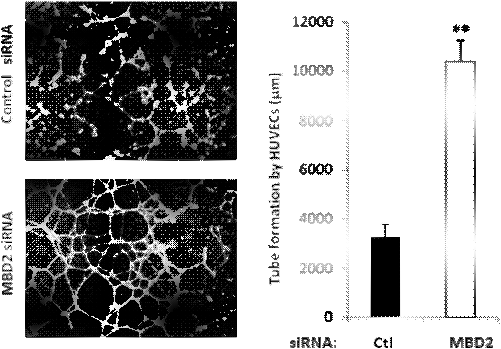
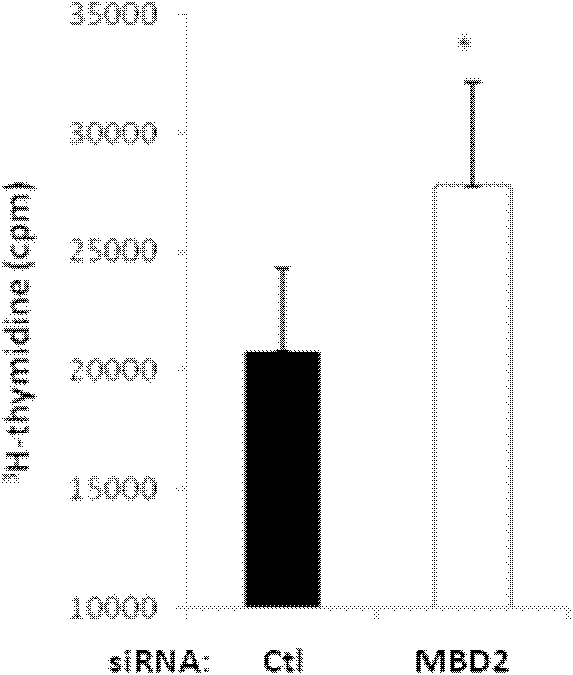
[0040] Example 2: MBD2 siRNA promotes angiogenesis in vitro and protects endothelial cells from H 2 o 2 induced apoptosis

[0041] To detect tube formation, Matrigel was placed in a 96-well plate (100 μL / well), at 37°C for at least 30 minutes, and then endothelial cells transfected with siRNA (100 nmol / L) for 24 hours were seeded in the 96-well plate (culture medium EBM-2 culture medium containing 2% fetal bovine serum), the density is 2×10 4 cells / well, in 5% CO 2 , Incubate in a 37°C incubator for 24 hours. The tube formation was checked under the light microscope every 4 hours, and it was found that typical tube formation could be observed in the MBD2 siRNA transfection group 4 hours after inoculation, but almost no tube formation was found in the control siRNA group; 24 hours later, the MBD2 siRNA transfection group The average tube length (10.4±0.8mm) of the stained group was significantly higher than that of the control group (3.3±0.5mm, Figure 2A ).

[0042] In o...

Embodiment 3

[0045] Example 3: The effect of MBD2 gene knockout on the recovery of mouse hindlimb perfusion was detected by hindlimb ischemia experiment

[0046] Establishment of the hindlimb ischemia model: After the mice were anesthetized, expose and separate the proximal end of the femoral artery at the left groin, tie two knots at the proximal end of the femoral artery with 7-0 silk thread, and cut the femoral artery from between the two knots with scissors. Suture the incision.

[0047]The expression of MBD2 in the ischemic site: the femoral artery of the left hind limb was taken at different time periods, and the protein was extracted for Western blot detection to observe the expression of MBD2. Simultaneously, ischemic tissue and contralateral non-ischemic tissue were embedded in OCT, sliced ​​(10 μm), and immunohistochemically stained to detect the localization of MBD2 in blood vessels.

[0048] Western blot experiments showed that MBD2 protein was not expressed in MBD2-deficient ...


PUM


Abstract

The invention discloses an inhibitor for inhibiting the expression of MBD2 and application thereof, and relates to an inhibitor capable of inhibiting the expression of methylated CpG binding domain protein 2 in vivio and in vitro. The inhibitor comprises a nucleic acid molecule, a genetic structure, an siRNA molecule and a compound containing one or more of the nucleic acid molecule, the genetic structure and the siRNA molecule, and can be introduced into mammalian cells to inhibit the expression of the MBD2. The inhibitor for inhibiting the expression of the MBD2 is an siRNA, which has a nucleotide sequence in which the sense strand is 5'-GCAAGAGCGAUGUCUACUA-3' and the antisense strand is 5'-UAGUAGACAUCGCUCUUGC-3', or of which the 3' end is provided with two suspended basic groups tt, and has a nucleotide sequence in which the sense strand is 5'-GCAAGAGCGAUGUCUACUAtt-3' and the antisense strand is 5'-UAGUAGACAUCGCUCUUGCtt-3'. The application of the inhibitor in preventing and treating related diseases of vascular endothelial cell dysfunction provides a novel option for preparing medicaments or preparations for preventing and treating the related diseases of the vascular endothelial cell dysfunction by taking the MBD2 as a target.

Description

technical field [0001] The invention belongs to the technical field of molecular biology and biomedicine, and specifically relates to an inhibitor and an its application. Background technique [0002] Vascular endothelial cells line the inner surface of the entire cardiovascular system. They not only regulate the transportation of nutrients, various bioactive molecules and blood cells, but also are an important self-balancing organ. Normal endothelial cells can regulate various physiological and In response to chemical signals, a group of autocrine and paracrine substances are released, such as nitric oxide (NO), cycloprostaglandin and endothelium-derived hyperpolarizing factor, etc., to regulate the tension of blood vessels, inhibit platelet aggregation, cell adhesion, smooth muscle Cell proliferation, which plays a key role in the regulation of vascular function, also plays an important role in vascular diseases and their initial defense. However, when endothelial cells ...

Claims


Application Information

Patent Timeline
IPC IPC(8): C12N15/113A61K48/00A61P9/00A61P9/10
Inventor 王从义余其林
Owner TONGJI HOSPITAL ATTACHED TO TONGJI MEDICAL COLLEGE HUAZHONG SCI TECH
Why Eureka
Try Eureka