Method for separating and purifying human amniotic fluid-derived mesenchymal stemcells

A technology for the separation and purification of mesenchymal stem cells, applied in the field of separation and purification of amniotic fluid mesenchymal stem cells, can solve the problems of high cost and complicated operation, and achieve the effect of convenient raw material acquisition, simple acquisition method and cost reduction

Inactive Publication Date: 2011-01-19
ZHEJIANG UNIV
View PDF0 Cites 2 Cited by
  • Summary
  • Abstract
  • Description
  • Claims
  • Application Information

AI Technical Summary

Problems solved by technology

[0004] The traditional isolation and purification of human amniotic fluid stem cells requires amniocentesis, cell adheren

Method used


Image

  • Method for separating and purifying human amniotic fluid-derived mesenchymal stemcells
  • Method for separating and purifying human amniotic fluid-derived mesenchymal stemcells
  • Method for separating and purifying human amniotic fluid-derived mesenchymal stemcells

Examples


Embodiment 1

[0023] Example 1: Isolation, purification and expansion of human amniotic fluid stem cells in the discarded supernatant of amniotic fluid culture for prenatal diagnosis

[0024] (1) Aseptically take mid-term amniotic fluid samples. The amniotic fluid comes from outpatients in the prenatal diagnosis clinic of the Obstetrics and Gynecology Hospital Affiliated to Zhejiang University. With my consent, the discarded supernatant is used for related research. 12ml of mid-term amniotic fluid samples are centrifuged at 1500rpm for 10min; Clear, add 1m cell culture medium II, resuspend the cell pellet, transfer to T25 culture flask for culture. After 7 days, the adherent cells were harvested for prenatal diagnosis, and the discarded supernatant was collected for subsequent use; the final concentration of the cell culture medium II was composed as follows: 80% (v / v) low-sugar DMEM+20% (v / v) FBS+ 4ng / ml bFGF+100U / ml penicillin+100U / ml streptomycin.

[0025] (2) Step (1) Discard the super...

Embodiment 2

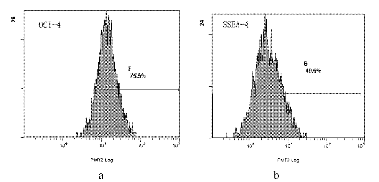
[0027] Example 2: Detection of Surface Antigen Characteristics, Immunogenicity and Totipotent Stem Cell Markers

[0028] The specific analysis method is as follows:

[0029] 1. Antigen characteristics and immunogenicity detection

[0030] (1) Digest the cells obtained in Example 1 with trypsin, add an equal amount of culture medium III, and terminate the digestion. The composition of the final concentration of the cell culture medium III is as follows: 85% (v / v) low-sugar DMEM+10% (v / v) FBS+100U / ml penicillin+100U / ml streptomycin;

[0031](2) After washing once with 10ml of PBS, resuspend in 1.2ml of PBS containing 1% (v / v) FBS to prepare a cell suspension, divide into 8 tubes according to the amount of 100 μL per tube, and control the amount of cells in each tube at 5×10 5 ~1×10 6 indivual;

[0032] (3) Add 20 μL each of CD29 and its isotype control, CD34 and its isotype control, CD105 and its isotype control, HLA-DR and its isotype control in sequence, and incubate at 4...

Embodiment 3

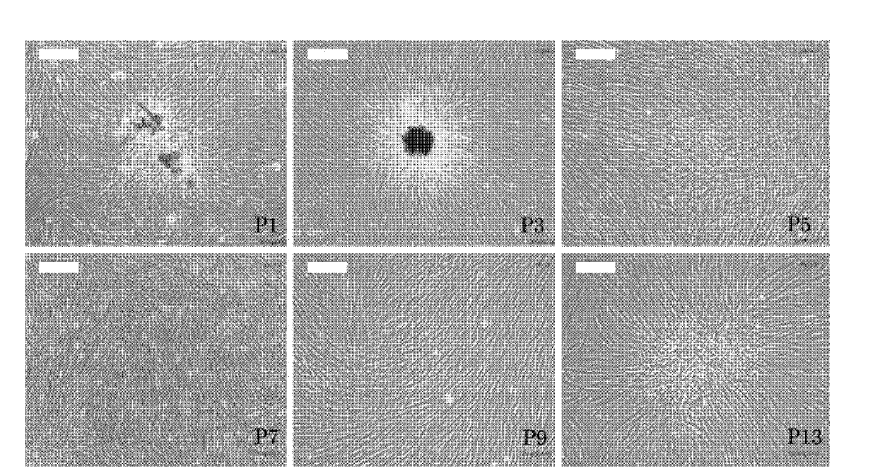
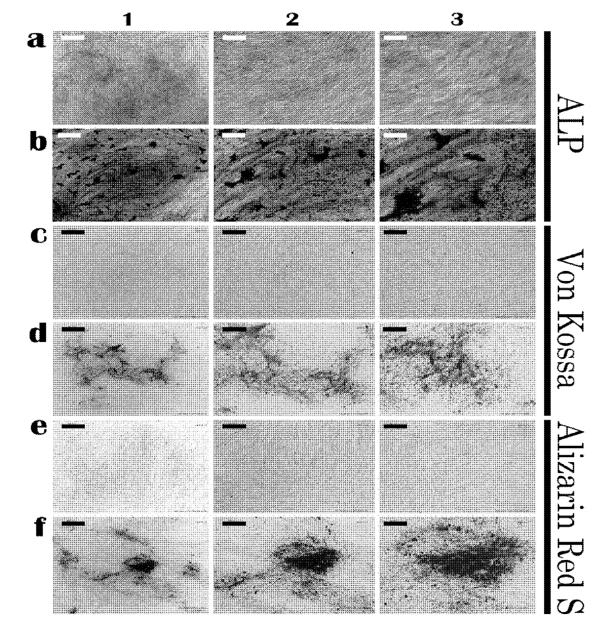
[0043] Example 3: Identification of multi-lineage differentiation potential

[0044] 1. Induced differentiation into adipocytes

[0045] Induction medium composition: low-sugar DMEM+15% (v / v) FBS+fat additive (1umol / L dexamethasone+10mg / L insulin+0.5mmol / L isobutylmethylxanthine+200umol / L indomethacin)

[0046] Induction method: Human amniotic fluid stem cells obtained in Example 1 were mixed with 3-5×10 3 piece / cm 2 The cell density was seeded in 24-well plates, and when 90% confluence was reached, it was replaced with adipose induction medium. The medium was changed every 3 to 4 days, and after 2 weeks of induction, positively stained fat particles were observed by Oil Red O staining ( Figure 4 ), and by RT-PCR (detection primers Forward: 5'-GCCATCCGCATCTTTCGA-3', Reverse: 5'-AGGACTCAGGGTGGTTCA-3'), the transcription factor peroxidase proliferator-activated receptor highly expressed in adipose tissue can be detected The expression of γ (PPARγ) indicates that human amnio...


PUM

PropertyMeasurementUnit
Diameteraaaaaaaaaa
Thicknessaaaaaaaaaa
Apertureaaaaaaaaaa

Abstract

The invention provides a method for separating and purifying human amniotic fluid-derived mesenchymal stemcells from discarded supernatant of prenatal diagnosis amniotic fluid culture. The cells are fibroblast-like amniotic fluid-derived mesenchymal stemcells obtained by extremely low-density cultivation in a specific culture medium, have multi-directional differentiation capability, have high amplification capability in comparison with adult cells and can maintain the original characteristics in 15 generations. The method has the main advantages that: (1) raw materials of the method are readily available and have no ethical tissues and an extremely high use value; (2) the cells obtained by using the method have high amplification capability, can maintain the characteristics in 15 generations and have the multi-directional differentiation capability; (3) the cell producing method of the invention is simple and high in efficiency, flow type and magnetic bead separation and purification processes and other separation and purification processes are not required, and the cost for obtaining the high-purity human amniotic fluid-derived mesenchymal stemcells is greatly reduced; and (4) the cells of the invention are applicable to tissue engineering, cell treatment and medicament screening and are suitable to be used as seeded cells of the tissue engineering.

Description

(1) Technical field [0001] The invention relates to a method for separating and purifying amniotic fluid-derived mesenchymal stem cells, in particular to a method for separating and purifying amniotic fluid mesenchymal stem cells derived from the discarded supernatant of amniotic fluid culture in prenatal diagnosis. (2) Background technology [0002] Human amniotic fluid-derived mesenchymal stem cells (Human amniotic fluid-derived mesenchymal stem cells, human amniotic fluid stem cells) are a type of fetal mesenchymal stem cells. In addition to the advantages of non-tumorigenicity and relatively easy culture of adult stem cells, they also have the following characteristics: 1 1. The source is rich and easy to obtain; 2. The proliferation ability and differentiation ability are much stronger than bone marrow-derived adult stem cells, and the differentiation ability is closer to embryonic stem cells. 3. It has low immunogenicity and has special application prospects in the res...

Claims


Application Information

Patent Timeline
IPC IPC(8): C12N5/0735
Inventor 黄荷凤徐晨明陈松长黄祎婷王金福胡文胜
Owner ZHEJIANG UNIV
Why Eureka
Try Eureka