Preparation method of silicon dioxide (SiO2) ultrasonic imaging radiography material

A technology of ultrasonic imaging and silicon dioxide, which is applied in the direction of echo/ultrasonic imaging agents, etc., to achieve the effects of pollution-free by-products, good biocompatibility, and affordable prices

Inactive Publication Date: 2011-05-25
SHANGHAI NORMAL UNIVERSITY
View PDF3 Cites 17 Cited by
  • Summary
  • Abstract
  • Description
  • Claims
  • Application Information

AI Technical Summary

Problems solved by technology

However, at present, the research on silica microspheres at home and abroad is mainly in the transportation of drugs or DNA and the release of drugs, while the application research in the field of ultrasound imaging is rarely reported, so we need to study it

Method used


Image

  • Preparation method of silicon dioxide (SiO2) ultrasonic imaging radiography material
  • Preparation method of silicon dioxide (SiO2) ultrasonic imaging radiography material
  • Preparation method of silicon dioxide (SiO2) ultrasonic imaging radiography material

Examples


Embodiment 1

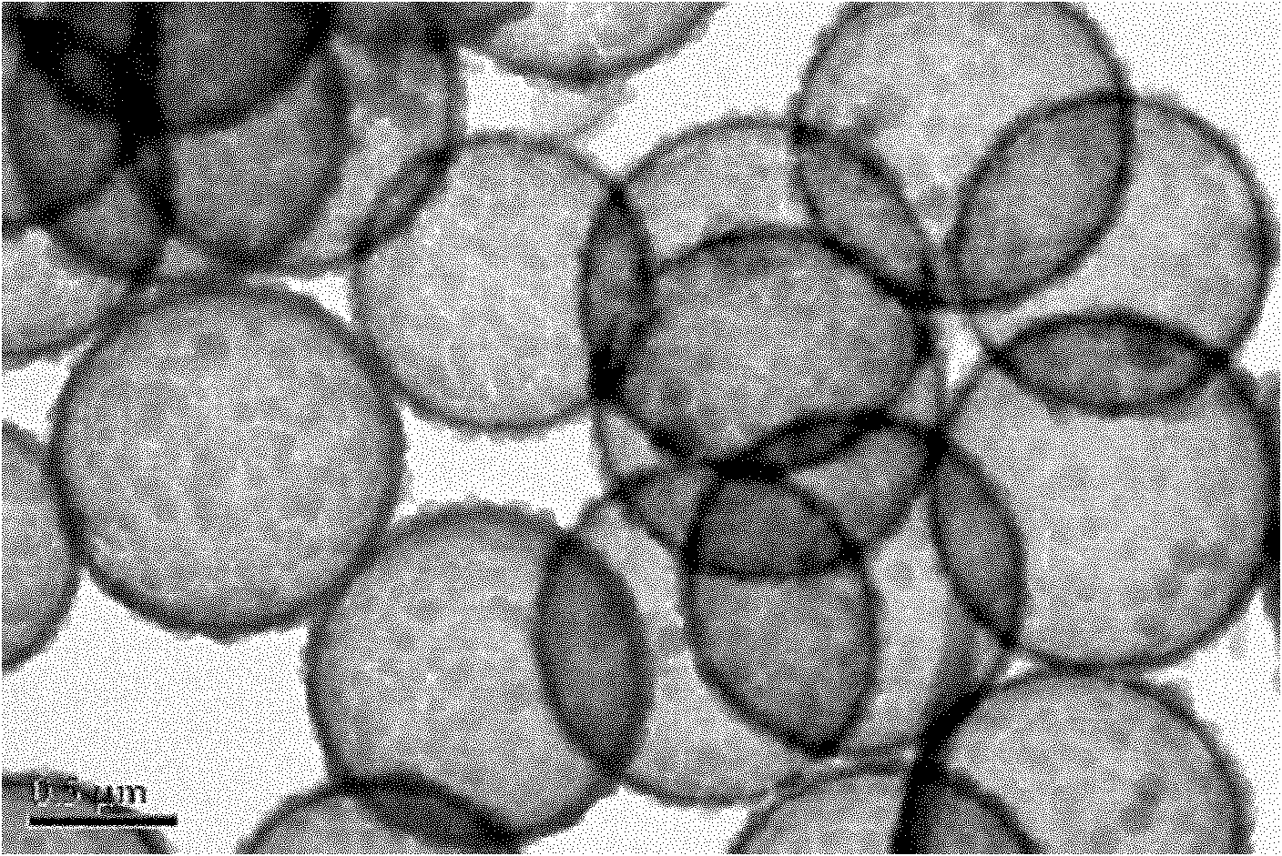
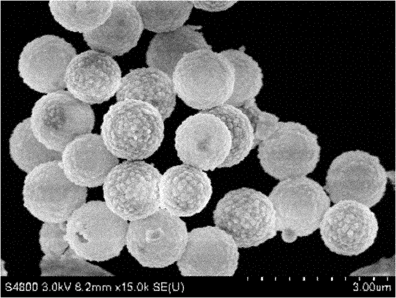
[0032] Measure 0.25 mL of commercial polystyrene microspheres (1 μm particle size) (5%, wt / v), add 7 mL of distilled water, 30 mL of isopropanol, 0.1 mL of tetraethyl orthosilicate (TEOS) (93 mg) , 0.01mL of 3-aminopropylsilane (9.5mg) and 0.2mL (mass concentration of 25% to 28%) ammonia water to adjust the pH value to 8 to 10, stir at room temperature for 3 hours, centrifuge, and wash to obtain the amino group on the surface. SiO 2 Microspheres.

[0033] Take 50mg of the above-mentioned SiO with amino groups on the surface 2 Microspheres, dispersed in 30mL tetrahydrofuran solution, extracted at 25°C for 8-10 hours, removed the hard template of polystyrene microspheres, and obtained SiO with amino groups on the surface 2 hollow microspheres.

[0034] According to the amino density test, the amino density is 8.65*10 -4 mol / g.

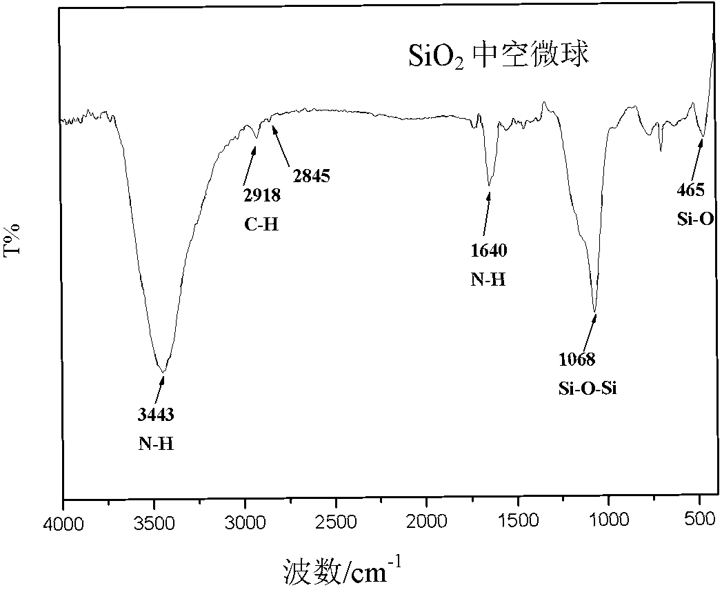
[0035] The transmission electron microscope picture is as figure 1 ; Field emission scanning electron microscope picture as figure 2 ; Infrared ...

Embodiment 2

[0037] The consumption of tetraethyl orthosilicate (TEOS) is 0.2ml (186mg), all the other are with embodiment 1. Infrared spectroscopy, transmission electron microscopy and field emission scanning electron microscopy detected that the prepared microspheres were SiO with amino groups on the surface. 2 hollow microspheres. According to the amino density test, the amino density is 4.85*10 -4 mol / g.

Embodiment 3

[0039] Take 6mg (3.46*10 -5 mol) PEG2000, dissolved in 20 mL of dimethyl sulfoxide (DMSO), added EDC (5 mg) and NHS (10 mg) according to the molar ratio of PEG: EDC: NHS = 1: 1: 2.5, stirred for 1 to 3 h, and used to activate the carboxyl group.

[0040] Add the SiO with amino group on the surface synthesized by 40mg embodiment 1 2 Hollow microspheres (the molar ratio of surface amino groups to PEG carboxyl groups is 1:1), slowly stirred at room temperature for 6-10 hours, washed, centrifuged, and freeze-dried to obtain very good biocompatibility, surface modified with PEG Alcoholic hollow SiO 2 microspheres for use in ultrasound imaging. The particle size is uniform, the average particle size is 960nm, and the thickness of the shell layer is suitable, which is 30-50nm.

[0041] field emission scanning electron microscope Figure 4 , it can be seen that there is almost no obvious change in its morphology after modification.

[0042] Infrared spectrum picture as Figure ...


PUM


Abstract

The invention relates to a preparation method of a silicon dioxide (SiO2) ultrasonic imaging radiography material. The preparation method comprises the following steps: hydrolyzing a non-functional silicon source and an amino-functional silicon source in a water-alcohol system by utilizing a hard template so as to obtain silicon spheres with amino on surfaces; removing the hard template by extraction to obtain hollow SiO2 spheres with amino on surfaces with uniform particle sizes; and carrying out surface modification by coupling the amino group on the surfaces of the SiO2 spheres with a carboxyl group in polyethylene glycol (PEG) so as to modify the PEG with good biocompatibility on the surfaces of the spheres, and finally obtaining the SiO2 ultrasonic imaging radiography material which has the advantages of good dispersibility, good biocompatibility, suitable cavity dimension and shell thickness and is applicable to ultrasonic imaging.

Description

technical field [0001] The invention relates to the field of ultrasound imaging contrast materials, in particular to a preparation method of silicon dioxide ultrasound imaging contrast materials. Background technique [0002] Contrast-enhanced ultrasound is the third revolution of ultrasound technology after two-dimensional gray-scale ultrasound and color Doppler ultrasound. Significant differences between them, thereby enhancing the display of organs or lesions. With the extension and development of molecular biology technology, the development of molecular imaging technology and the improvement of the performance of ultrasonic imaging equipment, contrast-enhanced ultrasound technology can effectively enhance the two-dimensional ultrasound imaging of various organs, reflecting the different blood of normal tissues and diseased tissues. The superiority of perfusion, contrast agent distribution and image dynamic changes in the diagnosis and differentiation of lesions has att...

Claims


Application Information

Patent Timeline
Patent Type & Authority Applications(China)
IPC IPC(8): A61K49/22
Inventor 杨仕平胡鹤周宏杨红李雪健刘国华
Owner SHANGHAI NORMAL UNIVERSITY
Why Eureka
Try Eureka