Method for simultaneously labelling collagen type IV, macrophage and neovascularisation of tumors

A technology for macrophages and new blood vessels, applied in the field of tissue immunofluorescence, can solve the problems of not being able to satisfy multiple staining, and achieve the effects of convenient histomorphological analysis, strong detection specificity, and high detection sensitivity

Inactive Publication Date: 2011-05-25
WUHAN UNIV
View PDF3 Cites 8 Cited by
  • Summary
  • Abstract
  • Description
  • Claims
  • Application Information

AI Technical Summary

Problems solved by technology

Therefore, ordinary organic fluorescent dyes cannot meet the needs of multiple staining

Method used


Image

  • Method for simultaneously labelling collagen type IV, macrophage and neovascularisation of tumors
  • Method for simultaneously labelling collagen type IV, macrophage and neovascularisation of tumors
  • Method for simultaneously labelling collagen type IV, macrophage and neovascularisation of tumors

Examples


Embodiment 1

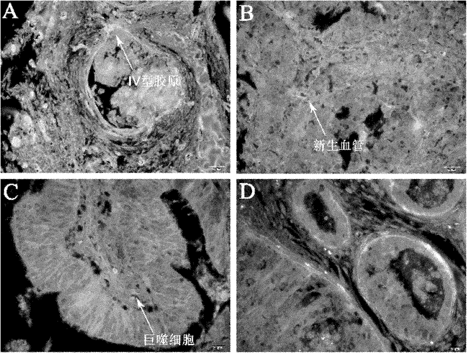
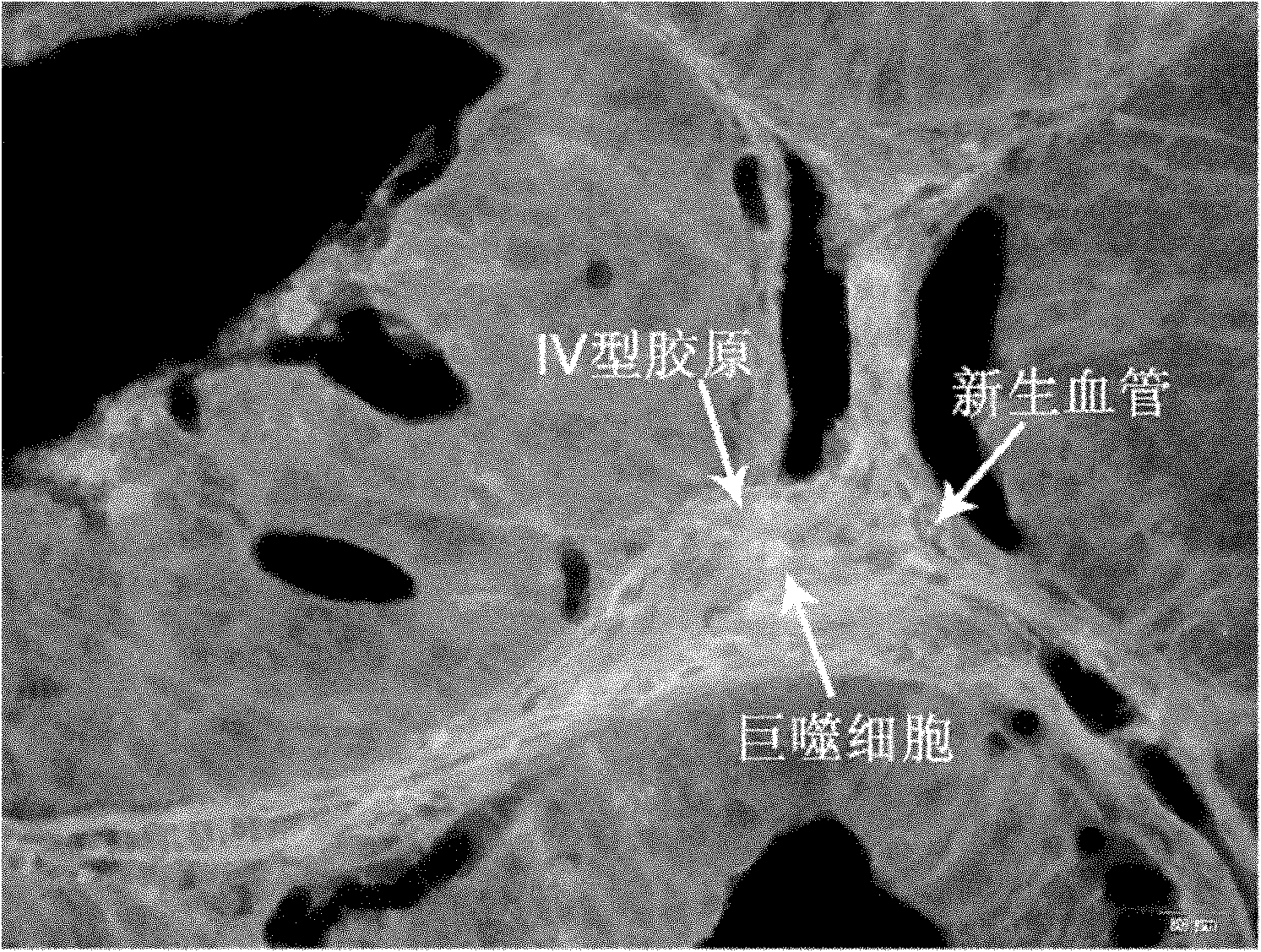
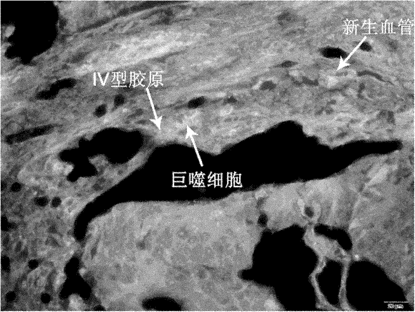
[0054] A method for simultaneously labeling tumor stroma type IV collagen, macrophages and angiogenesis, the steps of which are:

[0055] 1. Formalin-fixed, paraffin-embedded, 4 μm thick sections of gastric cancer tissue were fixed on poly-lysine-treated detachment-resistant glass slides.

[0056] 2. Preparation of tissue slices, put the tissue slices into xylene for dewaxing three times (in different vessels), 5 minutes each time, after dewaxing, put the tissue slices into absolute ethanol for 5 minutes, 95% alcohol for 2 minutes , 95% alcohol for 2 minutes and 80% alcohol for 2 minutes, rinse with running water for 3 to 5 minutes.

[0057] 3. For antigen retrieval, prepare trisodium citrate buffer with 29.41 g of trisodium citrate (Sinopharm Chemical Reagent Co., Ltd.), 1000 ml of double distilled water, 10.5 g of citric acid (Sinopharm Chemical Reagent Co., Ltd.), 500 ml of double distilled water ml to prepare citric acid buffer solution, respectively take 20.25ml of triso...


PUM

PropertyMeasurementUnit
diameteraaaaaaaaaa

Abstract

The invention discloses a method for simultaneously labelling collagen type IV, macrophage and neovascularisation of tumors. The method comprises the following steps: 1. immobilizing tissue sections on a plus microscope slide processed by polylysine; 2. dewaxing the tissue sections in dimethylbenzene; 3. carrying out antigen retrieval; 4. washing the sections; 5. carrying out confining; 6. addingprimary antibodies; 7. washing the sections; 8. carrying out confining in the same way as the step 5; 9. using the fragment antigen-binding F(ab')2-QDs525 of goat anti-mouse immunoglobulin G labelledby quantum dot QDs525, the fragment antigen-binding F(ab')2-QDs585 of goat anti-rabbit immunoglobulin G labelled by quantum dot QDs585 and the fragment antigen-binding F(ab')2-QDs655 of rabbit anti-goat immunoglobulin G labelled by quantum dot QDs655 as secondary antibodies and dropwise adding the mixture after removing the confining liquid; 10. washing the sections; and 11. sealing the sections:preparing buffered glycerol with glycerol and 10ml of tris buffered saline (TBS) and storing the sections after sealing the sections with the buffered glycerol. The method is used for efficiently, accurately, rapidly and simultaneously detecting various components in tumor tissue microenvironment.

Description

technical field [0001] The invention belongs to the technical field of tissue immunofluorescence, and more specifically relates to a quantum dot three-color fluorescent labeling method for detecting tumor interstitial type IV collagen, macrophages and tumor neovascularization, which is suitable for accurate and accurate detection of changes in human tumor tissue microenvironment components. Quantitative detection. Background technique [0002] The occurrence and development of tumors not only involve the tumor cells themselves, but also have a close relationship with the microenvironment around the tumor cells. The malignant tumor (tumor) microenvironment consists of tumor cells and their surrounding fibroblasts, epithelial cells, innate and specific immune cells, tumor blood vessels, mesenchymal cells and their expression products and metabolites. There are many components in the tumor microenvironment. Due to the lack of effective technology, the current research on the t...

Claims


Application Information

Patent Timeline
Patent Type & Authority Applications(China)
IPC IPC(8): G01N33/577G01N33/533
Inventor 彭春伟李雁庞代文朱小波
Owner WUHAN UNIV
Why Eureka
Try Eureka