Immunochromatographic test strip for quantitatively detecting troponin I in whole course and preparation method thereof

An immunochromatographic test strip, troponin technology, applied in biological tests, measuring devices, analytical materials, etc., to achieve accurate diagnosis of diseases and identification of infections, good labeling stability, and safe operation.

Inactive Publication Date: 2011-06-08
GUANGZHOU WONDFO BIOTECH
View PDF9 Cites 34 Cited by
  • Summary
  • Abstract
  • Description
  • Claims
  • Application Information

AI Technical Summary

Problems solved by technology

This technology has not yet been widely used in the field of disease diagnosis. It

Method used


Image

  • Immunochromatographic test strip for quantitatively detecting troponin I in whole course and preparation method thereof
  • Immunochromatographic test strip for quantitatively detecting troponin I in whole course and preparation method thereof

Examples


Example Embodiment

[0025] Example 1

[0026] In the embodiment of the present invention, the troponin I antibody used is a monoclonal antibody prepared by conventional monoclonal antibody technology, and the specimen is detected using the principle of the double antibody sandwich method to detect the troponin I antigen.

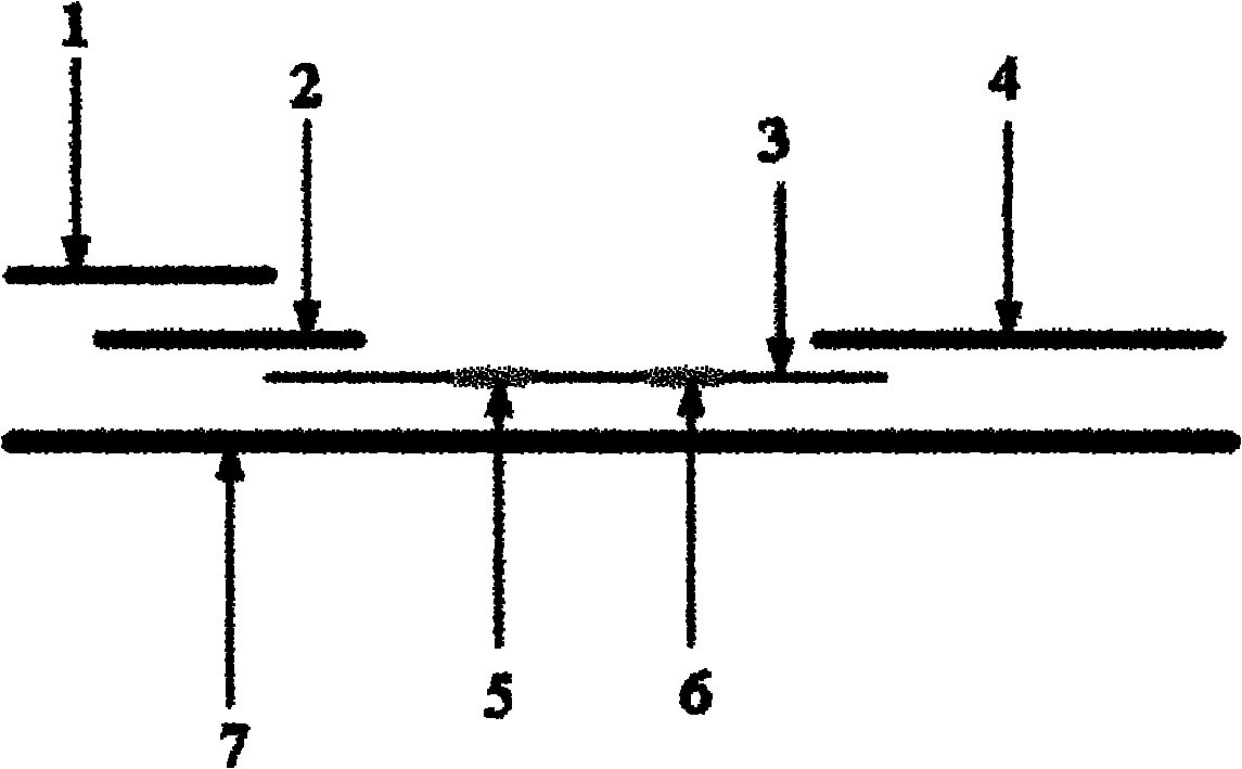
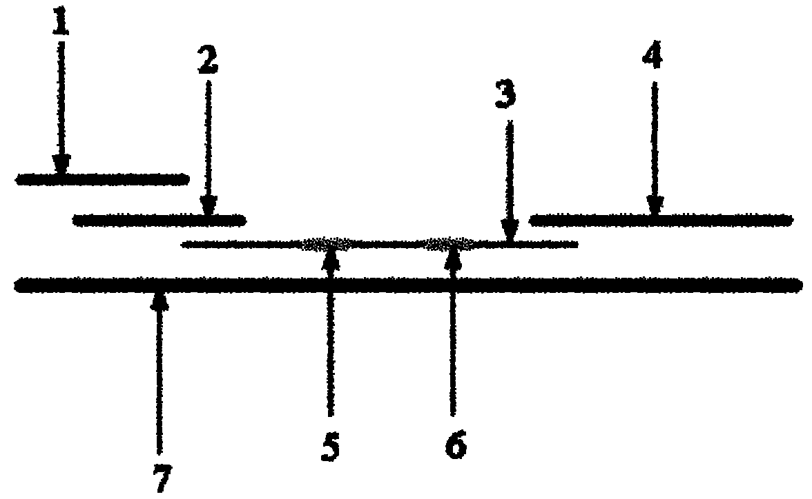
[0027] Such as figure 1 As shown, in this embodiment, the immunochromatographic test strip for quantitative detection of troponin I throughout the whole process includes a distal end and a proximal end. The sample pad 1 is located at the proximal end of the test strip, and it contains a hydrophilic hole. Diaphragm, sample pad 1 is the sample application area, which is used to draw samples for troponin I testing. Between the sample pad 1 and the distal end, a glass cellulose membrane marking pad 2, a nitrocellulose membrane coating membrane 3, and an absorbent paper 4 are sequentially overlapped. The sample pad 1, the glass fiber membrane marking pad 2, the nitrocellulose coating f...

Example Embodiment

[0038] Example 2

[0039] The structure of the test strip in this embodiment is the same as that of the first embodiment.

[0040] The concentration of the troponin I monoclonal antibody labeled with fluorescent latex particles is 0.1 mg / ml, and the spray film dosage on the test strip is 100 μl / 25 cm. The concentration of the troponin I monoclonal antibody coated in the detection area is 1 mg / ml, and the spray film dosage on the test strip is 100 μl / 25 cm. The concentration of the goat anti-mouse IgG is 1.5 mg / ml, and the spray film dosage on the test strip is 100 μl / 25 cm.

[0041] The preparation method of this embodiment except for the troponin I monoclonal antibody in step 2: the fluorescent latex is 15 μg / 100 μl. The rest are the same as in Example 1, and the method of use is also the same as in Example 1.

Example Embodiment

[0042] Example 3

[0043] The structure of the test strip in this embodiment is the same as that of the first embodiment.

[0044] The concentration of the troponin I monoclonal antibody labeled with fluorescent latex particles is 0.8 mg / ml, and the spray film dosage on the test strip is 100 μl / 28 cm. The concentration of the troponin I monoclonal antibody coated in the detection area is 2 mg / ml, and the spray film dosage on the test strip is 100 μl / 28 cm. The concentration of the goat anti-mouse IgG is 2 mg / ml, and the spray film dosage on the test strip is 100 μl / 28 cm.

[0045] The preparation method of this embodiment except for the troponin I monoclonal antibody in step 2: the fluorescent latex is 100 μg / 100 μl. The rest are the same as in Example 1, and the method of use is also the same as in Example 1.

[0046] In an embodiment of the present invention, the immunochromatographic test strip for quantitative detection of troponin I throughout the entire process is assembled in...


PUM

PropertyMeasurementUnit
Diameteraaaaaaaaaa
Wavelengthaaaaaaaaaa

Abstract

The invention discloses an immunochromatographic test strip for quantitatively detecting troponin I in the whole course. The test strip is formed by sticking a sample pad, a glass fiber membrane labeling pad, a nitrocellulose coating membrane and absorbent paper to a substrate in sequence through lap joint, wherein a fluorescent latex particle labeled troponin I monoclonal antibody is coated on the glass fiber membrane labeling pad; the nitrocellulose coating membrane comprises a detection region and a quality control region; the detection region is coated by another troponin I monoclonal antibody with epitope different from that of the fluorescent latex particle labeled troponin I monoclonal antibody; and the quality control region is coated by goat anti-mouse IgG (immunoglobulin G). The test strip has the advantages of safety, simpleness and convenience in operation, suitability for single human/sample detection, rapidness and the like and can sensitively and quantitatively determine the troponin I in the whole course in 10 seconds, diagnose the diseases and identify infection more rapidly and accurately and detect the infection conditions and determine the curative effects of antibiotics.

Description

Technical field [0001] The invention belongs to the field of medical testing, and in particular relates to an immunochromatographic test strip for quantitatively detecting troponin I in the whole process and a preparation method thereof. Background technique [0002] Cardiovascular diseases have a high incidence, high disability, high mortality, and many complications, which are extremely harmful to health. Cardiovascular testing is the "bottleneck" subject in the entire cardiovascular field. Only by correctly diagnosing the disease in the shortest possible period of time can it be possible to apply advanced treatment methods to the clinic in a timely and effective manner to benefit patients. With the deepening of research on cardiovascular diagnostic markers, the clinical significance of relevant markers has become more clear. The detection of cardiac markers can directly affect the clinical diagnosis, risk stratification, treatment options and prognosis judgments of patients wi...

Claims


Application Information

Patent Timeline
IPC IPC(8): G01N33/68G01N33/533G01N33/558G01N33/577
Inventor 王继华周楠
Owner GUANGZHOU WONDFO BIOTECH
Why Eureka
Try Eureka