Method for preparing contact lens-shaped amniotic dressing

A technology for contact lenses and amniotic membrane dressings, applied in dressings, viscous dressings, ophthalmic treatment, etc., can solve problems such as non-covering of amniotic membrane, displacement of amniotic membrane, and irritation of eyes by hard materials.

Inactive Publication Date: 2011-08-03
韩国百朗德株式会社
View PDF4 Cites 5 Cited by
  • Summary
  • Abstract
  • Description
  • Claims
  • Application Information

AI Technical Summary

Problems solved by technology

However, using Prokera TM The following problems can arise: i) the amnion does not cover the outer peripheral area of ​​the corneal protrusion; ii) the amnion may be displaced by blinking; iii) a portion of the amnion may be damaged by blinking before the epithelial cells grow; and iv) stiff material irritating to eyes

Method used


Image

  • Method for preparing contact lens-shaped amniotic dressing
  • Method for preparing contact lens-shaped amniotic dressing
  • Method for preparing contact lens-shaped amniotic dressing

Examples


Embodiment 1

[0049] Embodiment 1: the preparation of amniotic membrane

[0050] (1-1) Preparation of human amniotic membrane

[0051] Human amnion was processed by the method disclosed in the paper by Kim and Tseng (Kim JC and Tseng SC, Cornea, 14:473-84 (1995)). Isolation of amniotic membranes from placentas of pregnant women undergoing caesarean section. Pregnant women were prescreened for viral hepatitis B and C, HIV, and syphilis, and only those placentas with negative maternal blood serology were used. The amniotic membrane thus obtained was successively washed several times with sterile saline solution and sodium hypochlorite, and repeatedly washed with purified water. The obtained membrane is stored under low temperature conditions (2-8° C.) for future use.

[0052] (1-2) Preparation of bovine amniotic membrane

[0053] The bovine amnion was processed through the following steps. Bovine placentas were obtained after parturition, and the amniotic membranes were isolated therefro...

Embodiment 2

[0056] Embodiment 2: the preparation of mold and ring

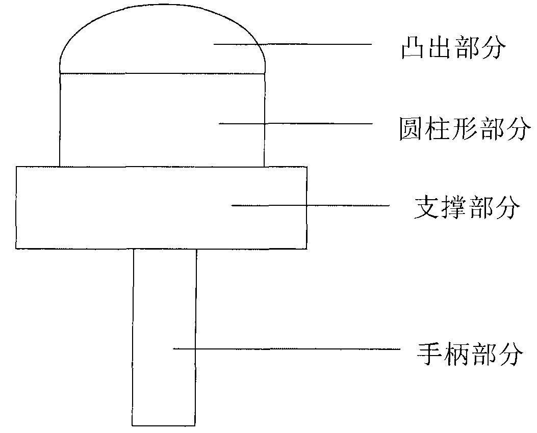
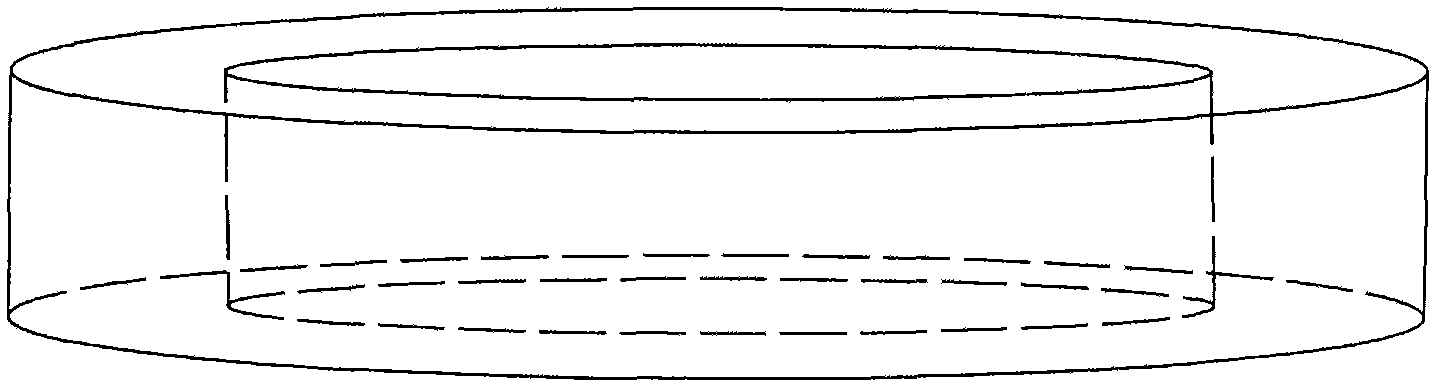
[0057] Such as figure 1 and 2 A mold made to form the amniotic membrane in the shape of a contact lens and a ring (or "O-ring") used to contact the amniotic membrane with the mold are shown. The mold consisted of a cylindrical part with a diameter of 14 mm, a convex part with a base arc of 8.4 to 8.8 mm, a support part and a handle part. The supporting part includes a supporter supporting a ring for contacting and fastening the amniotic membrane, and the handle part has a cylindrical shape connected to the middle of the bottom surface of the supporting part. The mold is made of acetal resin. manufacture figure 2 O-rings to fit each die with a specific base arc. O-rings were made of Teflon (14.1 mm internal diameter) or perfluoroelastomer (12 mm internal diameter).

Embodiment 3

[0058] Example 3: Preparation of Amniotic Membrane

[0059] In order to make the amniotic membrane obtained in (1-1) to (1-3) have a contact lens shape, the amniotic membrane was brought into contact with the contact lens-shaped mold obtained in Example 2, and then fixed on the mold with an O-ring.

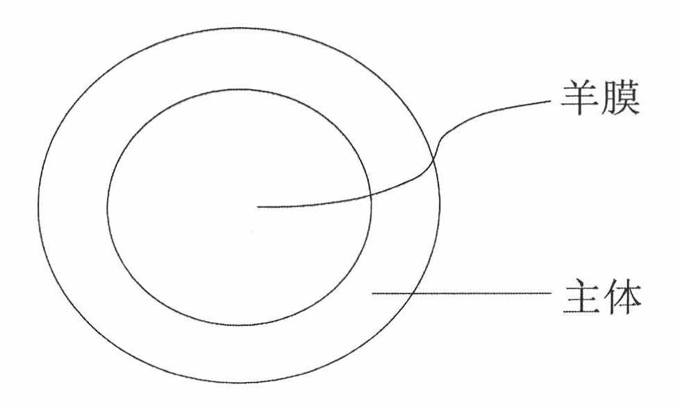
[0060] Specifically, the amniotic membrane was cut to the size of the mold, and then its epithelial layer was in contact with the convex part of the mold ( figure 1 ). In the case of overlapping amniotic membranes, overlap the last layer of amniotic membrane with its epithelial layer facing outwards. Then the O-ring ( figure 2 ) placed on the amniotic membrane or overlapping amniotic membranes to completely cover the mold, and then dried naturally, freeze-dried or vacuum-dried in a clean bench. Top perspective view of dried amniotic membrane (including mold) as image 3 shown.


PUM

PropertyMeasurementUnit
diameteraaaaaaaaaa
thicknessaaaaaaaaaa
diameteraaaaaaaaaa

Abstract

The present invention relates to a method for preparing a contact lens-shaped amniotic dressing and a contact lens-shaped amniotic dressing prepared therefrom for treating ocular surface diseases, which does not require the use of sutures or an adhesion material. The inventive contact lens-shaped amniotic dressing is capable of solving the problems associated with suturing an amniotic membrane, e.g., highly delicate surgical techniques of suturing, long surgery time, stitch abscess, granuloma formation, tissue necrosis, and discomfort of patients; and the problems associated with the use of a support, e.g., the elimination of the support by eye blinking, breaking of the support, and discomfort.

Description

technical field [0001] The present invention relates to a method for the preparation of a contact lens-like amniotic dressing for the treatment of diseases of the ocular surface which does not require the use of sutures or adhesive materials. Background technique [0002] The amniotic membrane is a membrane with a thickness of 0.02 to 0.5 mm located in the inner surface of the placenta, which has a stacked structure of epithelial cells, basement membrane, compact layer, fibroblast layer, and spongy layer. The amniotic membrane is avascular and does not express histocompatibility antigens, so immune rejection does not occur after its transplantation (Akle CA et al., Lancet, 2: 1003-5 (1981); Dua HS et al., Br. J. Ophthalmol. , 83:748-52 (1999)). Since the basement membrane of the amniotic membrane is composed of type IV collagen, laminin, and α6 / β4 integrin, and its interstitial tissue is composed of type I collagen, type III collagen, hyaluronic acid, fibronectin, and α5 / β1...

Claims


Application Information

Patent Timeline
Patent Type & Authority Applications(China)
IPC IPC(8): A61F13/02A61F9/00A61L15/40
CPCA61K35/48B29C63/02B29D11/00038A61L27/3604A61F9/0017A61L2/081
Inventor 梁恩敬丁三炫金成胞朱址熏李钟源洪钟明金在灿全然婌李性春白光一
Owner 韩国百朗德株式会社
Why Eureka
Try Eureka