Sca-1+/CD34- uterine stem cells and separation method thereof

A CD34-, separation method technology, applied in the field of stem cells, can solve the problem of the treatment mechanism and effect of uterine stem cell ischemic diseases, which have not been reported.

Inactive Publication Date: 2011-11-02
SHANXI MEDICAL UNIV
View PDF3 Cites 8 Cited by
  • Summary
  • Abstract
  • Description
  • Claims
  • Application Information

AI Technical Summary

Problems solved by technology

In addition, there is no report on the mechanism and effect of this kind of uterine stem cells in angiogenesis and the treatment of ischemic diseases.

Method used


Image

  • Sca-1+/CD34- uterine stem cells and separation method thereof
  • Sca-1+/CD34- uterine stem cells and separation method thereof
  • Sca-1+/CD34- uterine stem cells and separation method thereof

Examples


Embodiment 1

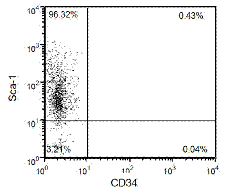
[0026] Example 1: Sca-1 + / CD34 - Isolation of Uterine Stem Cells 。

[0027] 1) 8-10 week old mice are anesthetized with isoflurane, the trachea is intubated, the ventilator maintains breathing, and 2-3% isoflurane maintains the anesthesia.

[0028] 2) The anesthetized mouse is in the supine position, the chest is opened in the center, the heart bag is torn, the right atrium is cut, and 0.9% saline is continuously infused from the aorta with physiological pressure, and the tissue is flushed until it flows out from the right atrium (no blood contamination) ) Of saline.

[0029] 3) Open the abdomen, take out the uterus, and chop it fully.

[0030] 4) Digest with 0.25% trypsin, 2mg / ml collagenase, 0.01% DNase at 37°C for 1 hour to minimize the damage of cell surface markers.

[0031] 5) From the above digested tissues, use magnetic bead-labeled anti-CD34 antibody (stemcell, Canada) to screen out CD34 by magnetic bead screening - cell. The step is to collect the digestion fluid, filter th...

Embodiment 2

[0035] Example 2: Sca-1 + / CD34 - Cultivation of Uterine Stem Cells.

[0036] The cells selected in Example 1 were inoculated into 35mm culture plates, and 1% methylcellulose basal medium, 10% fetal bovine serum, 4.5×10 -4 M thioglycerol, 25μg / mL ascorbic acid, 2mM glutamine, 200μg / mL saturated transferrin, the culture environment is kept at humidity ≥95%, temperature 37℃, CO 2 Concentration 5%.

Embodiment 3

[0037] Example 3: Sca-1 + / CD34 - Differentiation of Uterine Stem Cells.

[0038] Differentiation medium composition: IMDM medium, 10% fetal bovine serum and 25% primary endothelial cell culture medium.

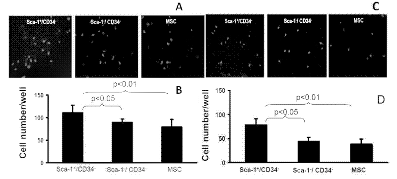
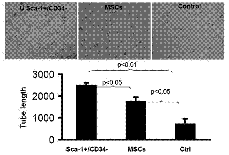
[0039] Sca-1 + / CD34 - Uterine stem cells, Sca-1 - / CD34 - Cells and bone marrow mesenchymal stem cells (MSC) cell extracts were added to Matrigel, and human umbilical cord blood endothelial cells (HUVEC) and smooth muscle cells (SMC) were cultured separately, and cultured in normal medium as a control group. The length of the formed new blood vessels is used to evaluate the ability of HUVECs to form vascular structures.

[0040] The results show that Sca-1 + / CD34 - Uterine stem cells are more capable of inducing endothelial cells to form vascular structures than Sca-1 - / CD34 - And bone marrow MSC are strong, see specifically figure 2 , image 3 .

[0041] figure 2 A-B shows Sca-1 + / CD34 - Uterine stem cells induce the migration of human umbilical cord vascular endothelial cells...


PUM


Abstract

The invention discloses Sca-1+ / CD34- uterine stem cells, which are separated from the uterus of a mammal. In the invention, the CD34 and Sca-1 are expressed in form of CD34- and Sca-1+. The separating method of the Sca-1+ / CD34- uterine stem cells comprises: screening CD34- cells from digested uterine tissues by using magnetic bead marked antibody against CD34; and separating Sca-1+ cells from CD34- cells by using magnetic bead marked antibody against Sca-1, and obtaining Sca-1+ / CD34- uterine stem cells. Experiments prove that the Sca-1+ / CD34- uterine stem cells are capable of inducing endothelial cells to form vascular structures and forming lots of functional blood vessels, and can restore tissue perfusion to a certain degree and reduce tissue damage; and in-vivo level proves the Sca-1+ / CD34- uterine stem cells have treatment effect on ischemic diseases and can be used in preparation of medicines for preventing or treating ischemic diseases.

Description

technical field [0001] The present invention relates to a stem cell, in particular to a stem cell extracted from the uterus. The present invention also relates to the separation method of the uterine stem cells and the application of the uterine stem cells. Background technique [0002] With the change of people's lifestyle, ischemic diseases have become one of the diseases with the highest incidence in the world in recent years, and the incidence has been rising. Ischemic disease is a disease that causes poor tissue perfusion due to vascular stenosis or occlusion, and eventually causes organ dysfunction or even failure. disease, cerebral infarction, etc. Under normal conditions, angiogenesis is the body's own compensatory mechanism for ischemia and hypoxic injury. In the early stage of myocardial ischemia, capillary density increases compensatoryly, and new blood vessels are formed around blocked or narrowed coronary arteries to establish collateral circulation, thereby ...

Claims


Application Information

Patent Timeline
Patent Type & Authority Applications(China)
IPC IPC(8): C12N5/074A61K35/48A61P9/10
Inventor 李仁科解军刘志贞孙卓
Owner SHANXI MEDICAL UNIV
Why Eureka
Try Eureka