Enzyme-labeling-liquid-based cytology staining kit for screening bladder cancer

A dyeing reagent and cytology technology, which is applied in the field of enzyme-labeled liquid-based cytology technology to screen bladder tumors, can solve the problems of heavy workload and high false negative rate, and achieve the effects of easy interpretation, clear shape, and maximized cell information

Active Publication Date: 2011-11-30
合肥科久盛生物医药有限公司
View PDF9 Cites 9 Cited by
  • Summary
  • Abstract
  • Description
  • Claims
  • Application Information

AI Technical Summary

Problems solved by technology

[0006] The current HE staining methods are liquid-based thin-layer cell preparation method and traditional smear. The above methods rely solely on doctors to g

Method used


Image

  • Enzyme-labeling-liquid-based cytology staining kit for screening bladder cancer
  • Enzyme-labeling-liquid-based cytology staining kit for screening bladder cancer
  • Enzyme-labeling-liquid-based cytology staining kit for screening bladder cancer

Examples


Embodiment 1

[0041] 1. Enzyme-labeled liquid-based cytology staining kit components:

[0042] (1) Fixative: 10% formalin;

[0043] (2) ELISA liquid-based cytological staining solution: 2-30.5g / l naphthol AS-BI phosphate dissolved in dimethylformamide;

[0044] (3) Fuchsin hydrochloride: 1-10% fuchsin hydrochloride;

[0045] (4) Sodium nitrite: 0.1-0.5M NaNO 2 ;

[0046] (5) Sodium acetate solution: 1.5-4.5M, pH 4.5-5.9;

[0047] (6) Hematoxylin counterstaining solution;

[0048] (7) filter paper

[0049] 2. Preservation solution composition:

[0050] (1) 45% methanol;

[0051] (2) 0.2-0.5mM EDTA;

[0052] (3) 0.1mM NaCl;

[0053] (4) 0.1M acetate buffer, pH 4-6;

[0054] (5)dd H 2 O.

[0055] 3. Sedimentation buffer composition:

[0056] Recipe a:

[0057] 0.5XPBS, pH=7.0-7.4, 2-5mM EDTA;

[0058] Recipe b:

[0059] 10mM Tris-HCl pH=7.0-7.4, 2-5mM EDTA, 0.9% (m / v) NaCl.

[0060] 3. Sampling and operation

[0061] A. Conventional production

[0062] Collect 15 mL of mid-m...

Embodiment 2

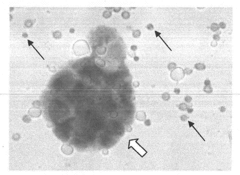
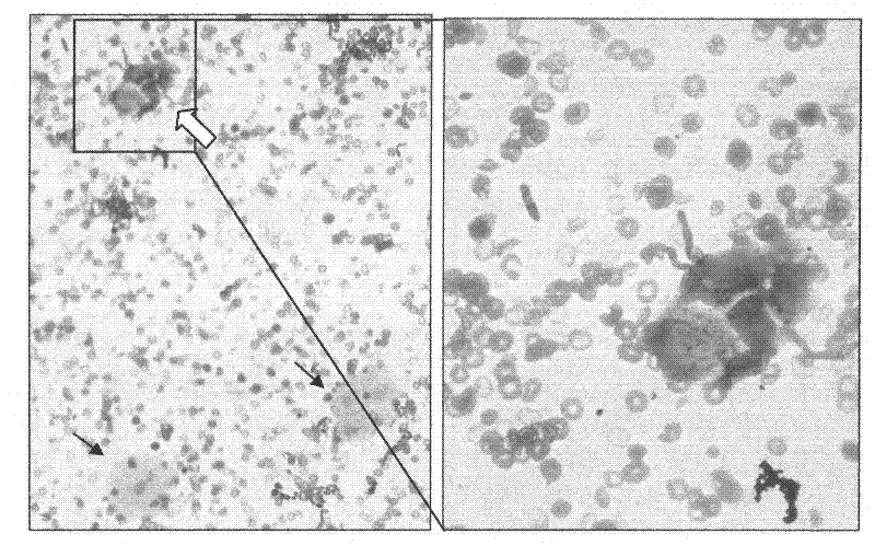
[0075] Three cases of patients and healthy persons who have been clinically identified as bladder cancer, liver cancer, lung cancer, colorectal cancer, gastric cancer, and cervical cancer were selected. Urine samples were taken according to the method in Example 1, and enzyme-labeled liquid-based cytological staining was performed. The results showed that all three patients with bladder cancer had Figure 1-3 The cytoplasm in the urine was stained red, but no cytoplasm red stained in the urine samples of other cancer patients and healthy people.

[0076] The above test results show that the high expression of acid phosphatase detected in urine samples is highly specific to bladder cancer, and the method and kit of the present application can be used for screening diagnosis or assisting screening and diagnosis of bladder cancer patients.

Embodiment 3

[0077] Example 3 Effect test of enzyme-labeled liquid-based cytology preservation solution and sedimentation buffer

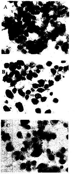
[0078] The bladder cancer cell line BIU-87 was preserved in the enzyme-labeled liquid-based cytological preservation solution described in this application and an existing preservation solution (purchased from Xiamen Maiwei Biotechnology Co., Ltd.) for 7 days, and then carried out with the sedimentation buffer. Film preparation and staining, the results obtained are as attached Figure 5 Shown, the preparation and staining effect ( Figure 5 A, 5B) The effect is significantly better than that of the control group cell preservation solution and sedimentation buffer (Xiamen Maiwei Biotechnology Co., Ltd., Figure 5 C). The specific comparison results are shown in the following table (more "+" means better effect):

[0079]

[0080] From this result, it can be seen that the enzyme-labeled liquid-based cytology preservation solution and the sedimentation buff...


PUM


Abstract

The invention relates to application of an enzyme-labeling-liquid-based cytology technology to the screening of bladder cancer. In the technology, a cell sample which is tabletted by a liquid-based tabletting method is treated by an acid phosphatase chemical staining method, and a bright red precipitate is formed in cytoplasm of abnormal cells by specific staining to ensure that the abnormal cells have remarkable marks. The invention provides a kit for the detection. The kit mainly comprises an enzyme-labeling-liquid-based cytology staining reagent, enzyme-labeling-liquid-based cytology preserving fluid and settlement buffer solution, and can provide high detection specificity and a good staining effect.

Description

technical field [0001] The invention relates to a detection kit for bladder cancer and its application, more precisely, it uses enzyme-labeled liquid-based cytology technology to screen bladder tumors. This technology uses acid phosphatase chemical staining method to process the cell samples prepared by liquid-based film preparation method, and uses this specific staining to form a bright red precipitate in the cytoplasm of abnormal cells, so that the abnormal cells are clearly marked. Background technique [0002] Bladder cancer is the most common urological tumor in my country, and about 90% of bladder cancer is bladder urothelial carcinoma. Bladder cancer has a high recurrence rate, 60% to 70% of patients may relapse, and 11% of relapsed patients may develop into invasive tumors. Currently, the diagnosis and follow-up of bladder cancer mainly rely on cystoscopy and urine cytology. The former is the gold standard for diagnosing bladder cancer, but it is an invasive test ...

Claims


Application Information

Patent Timeline
IPC IPC(8): C12Q1/42C12Q1/04
Inventor 沈玉先李俊田添王阳
Owner 合肥科久盛生物医药有限公司
Why Eureka
Try Eureka