Preparation method of drug carried microsphere capable of being positioning injected into tumor cavity
A technology of drug-loaded microspheres and polylactic acid, which is applied in anti-tumor drugs, pharmaceutical formulas, medical preparations of non-active ingredients, etc.
- Summary
- Abstract
- Description
- Claims
- Application Information
AI Technical Summary
Problems solved by technology
Method used
Image

Examples
Embodiment 1
[0026] (1) Preparation of drug-loaded microspheres that can be injected into tumor cavity
[0027] 1) Accurately weigh 8.0 mg of polylactic acid and 2.0 mg of paclitaxel and place in a vial, dissolve with 1.0 ml of acetone, stir well and set aside. Add 1mg of BSA to 10ml of water under constant stirring;
[0028] 2) Take 10.0ml of deionized water and put it in a 25ml beaker, put it into a large magnetic stirrer, start stirring and adjust the speed, so that the magnet rotates smoothly without hitting the wall, and the liquid surface is in a very good spindle shape;
[0029] 3) Use a 1ml disposable syringe to suck out the acetone solution and inject it into the beaker at one time. The injection should be slow and even, and the injection needle should be below the liquid level;
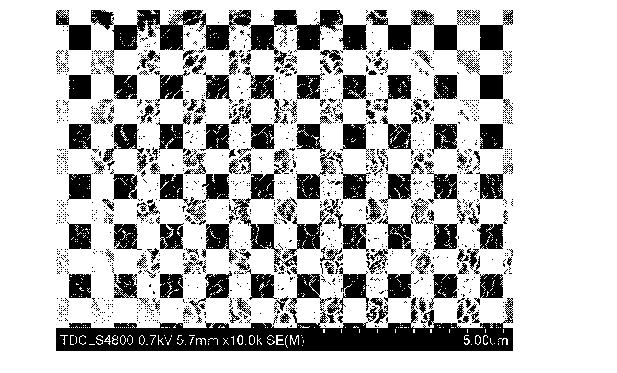
[0030] 4) After stirring for 3 hours, until the acetone is completely volatilized, an opalescent suspension is obtained, and freeze-dried to obtain white PLA drug-loaded nanoparticle powder.
[0031] 5...
Embodiment 2
[0033] 1) Accurately weigh 9.0mg of polylactic acid and 1.0mg of paclitaxel in a vial, dissolve with 1.0ml of acetone, stir well and set aside. Add 1mg of BSA to 10ml of water under constant stirring;
[0034] 2) Take 10.0ml of deionized water and put it in a 25ml beaker, put it into a large magnetic stirrer, start stirring and adjust the speed, so that the magnet rotates smoothly without hitting the wall, and the liquid surface is in a very good spindle shape;
[0035] 3) Use a 1ml disposable syringe to suck out the acetone solution and inject it into the beaker at one time. The injection should be slow and even, and the injection needle should be below the liquid level;
[0036] 4) After stirring for 3 hours, until the acetone is completely volatilized, an opalescent suspension is obtained, and freeze-dried to obtain white PLA drug-loaded nanoparticle powder.
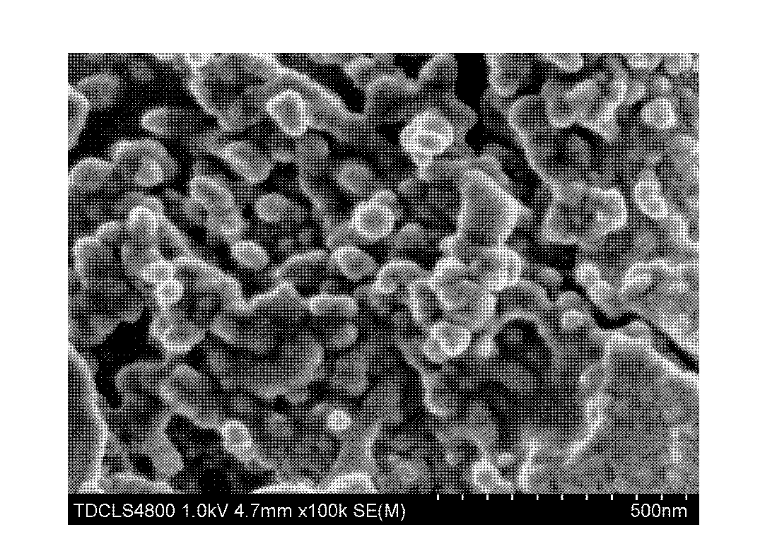
[0037] 5) Example 2 obtains the nano drug-loaded microspheres, observes the scanning electron microscope picture o...
Embodiment 3
[0039] 1) Accurately weigh 8.0 mg of polylactic acid and 2.0 mg of paclitaxel and place in a vial, dissolve with 1.0 ml of acetone, stir well and set aside. Add 2mg of BSA to 10ml of water under constant stirring;
[0040] 2) Take 10.0ml of deionized water and put it in a 25ml beaker, put it into a large magnetic stirrer, start stirring and adjust the speed, so that the magnet rotates smoothly without hitting the wall, and the liquid surface is in a very good spindle shape;
[0041] 3) Use a 1ml disposable syringe to suck out the acetone solution and inject it into the beaker at one time. The injection should be slow and even, and the injection needle should be below the liquid level;
[0042] 4) After stirring for 3 hours, until the acetone is completely volatilized, an opalescent suspension is obtained, and freeze-dried to obtain white PLA drug-loaded nanoparticle powder.
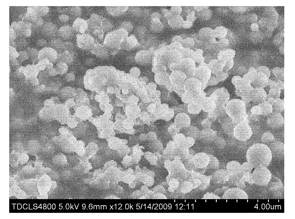
[0043] 5) The nano drug-loaded microspheres obtained in Example 3, after freeze-drying, observe the s...
PUM
| Property | Measurement | Unit |
|---|---|---|
| Particle size | aaaaa | aaaaa |
| Particle size | aaaaa | aaaaa |
Abstract
Description
Claims
Application Information
 - R&D
- Intellectual Property
- Life Sciences
- Materials
- Tech Scout
- Unparalleled Data Quality
- Higher Quality Content
- 60% Fewer Hallucinations
Browse by: Latest US Patents, China's latest patents, Technical Efficacy Thesaurus, Application Domain, Technology Topic, Popular Technical Reports.
© 2025 PatSnap. All rights reserved.Legal|Privacy policy|Modern Slavery Act Transparency Statement|Sitemap|About US| Contact US: help@patsnap.com