In vitro culture method for inducing human umbilical cord blood cells CD34+ to differentiate into Langerhans cell

A technology of cell differentiation and in vitro culture, applied in artificial cell constructs, blood/immune system cells, animal cells, etc.

Inactive Publication Date: 2012-08-15
THE FIRST AFFILIATED HOSPITAL OF THIRD MILITARY MEDICAL UNIVERSITY OF PLA
View PDF1 Cites 1 Cited by
  • Summary
  • Abstract
  • Description
  • Claims
  • Application Information

AI Technical Summary

Problems solved by technology

[0006] It has not been reported that by culturing them in a medium containing GM-CSF, TNE-α, SCF and Flt3L, cord blood CD34+ cells can be differentiat

Method used


Image

  • In vitro culture method for inducing human umbilical cord blood cells CD34+ to differentiate into Langerhans cell
  • In vitro culture method for inducing human umbilical cord blood cells CD34+ to differentiate into Langerhans cell
  • In vitro culture method for inducing human umbilical cord blood cells CD34+ to differentiate into Langerhans cell

Examples


Embodiment 1

[0022] Example 1 Isolation of CD34+ cells in umbilical cord blood

[0023] 1. Isolation of human cord blood mononuclear cells:

[0024] ①In the dark, add 10mL of human lymphocyte separation medium (density 1.077) into a 15mL centrifuge tube, carefully add 5mL of cord blood to the lymphocyte separation medium, and form a clear interface.

[0025] ②At room temperature, centrifuge horizontally at 400g (2500rmp, r=14.5 cm) for 25min.

[0026] ③ At this time, five layers can be seen in the centrifuge tube: the uppermost layer is plasma, the white misty layer between the plasma layer and the lymphocyte separation liquid is the mononuclear cell layer, and the white film between the lymphocyte separation liquid layer and the lowermost red blood cells The layer is the granulocyte layer.

[0027] ④ Suck off the uppermost layer of plasma, collect the mononuclear cells at the interface between the plasma layer and the lymphocyte separation medium, and try to suck out all the mononuclea...

Embodiment 2

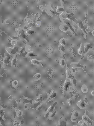
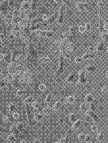
[0042] Example 2 Induction and production of Langerhans cells in vitro

[0043] steps:

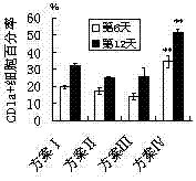
[0044] 1. Count the CD34+ cells obtained by magnetic bead sorting, and count them according to 1×10 4 cells / cm 2 Inoculated in 24-well plates, cultured with RPMI1640 complete medium plus the cytokines of the following four schemes, see Table 1. The concentration of each factor in the table is the final concentration of each factor in each mL of RPMI1640 medium.

[0045] Table 1 The scheme of inducing Langerhans cells in vitro with cytokine combination

[0046]

[0047] 2. On the first day after the separation of CD34+ cells, add RPMI-1640 culture medium containing 10% fetal bovine serum containing the cytokines in the table according to the scheme in Table 1, at 37°C, 5% CO 2 Culture; change the culture medium every 3 days. Two wells of 24-well plates were cultured in each protocol.

[0048] 3. The phenotype of Langerhans cells (CD1a) was detected by flow cytometry on the 6th day...

Embodiment 3

[0052] Example 3 Detecting the phenotypes of antigen-presenting molecules (HLA-DR) and co-stimulatory molecules (CD80, CD40) of Langerhans cells by flow cytometry

[0053] step:

[0054] 1. Digest the Langerhans cells obtained in Example 2 with trypsin, centrifuge at 200 g for 10 min (1000 rpm, r=14.5 cm), discard the supernatant, and obtain the Langerhans cell pellet.

[0055] 2. Add 1mL ice-cold PBS buffer to resuspend the cell pellet, and count the cells.

[0056] 3. According to 10 5 Cells / 100 μL concentration was adjusted for cell concentration, and 100 μL of Langerhans cell suspension was taken from each tube.

[0057] 4. Label PE / CY5-HLA-DR, FITC-CD80, FITC-CD40 monoclonal antibodies and corresponding isotype controls respectively, mix well, and incubate at 4°C for 40 minutes in the dark.

[0058] 5. Add 100 μL PBS to each tube, and use flow cytometer for fluorescence detection on the same day. Or fix the cells and perform flow cytometry within 5 days: add 500 μL of...


PUM


Abstract

An in vitro culture method for inducing human umbilical cord blood cells CD34+ to differentiate into Langerhans cell includes culturing human umbilical cord blood cells CD34+ in a medium containing GM-CSF (granulocyte-macrophage colony stimulating factor), TNF-alpha (tumor necrosis factor-alpha), SCF (stem cell factor) and Flt3L (FMS-like tyrosine kinase 3 ligand), and differentiating the human umbilical cord blood cells CD34+ into Langerhans cells. The Langerhans cells generated are highly pure and high in yield, and have the functions of recognizing, absorbing and processing antigens and the function of activating immune response.

Description

technical field [0001] The present invention relates to the development of a culture medium containing granulocyte-macrophage colony-stimulating factor (GM-CSF), tumor necrosis factor alpha (TNF-α), stem cell factor (SCF) and FMS-like tyrosinase 3 ligand (Flt3L) In vitro induction method for culturing umbilical cord blood CD34+ cells and making them differentiate Langerhans cells, and the resulting Langerhans cells have the characteristics of high purity and large yield, and have the ability to recognize, uptake, process and process antigens ability to initiate an immune response. Background technique [0002] Stem cells have the ability to divide indefinitely in a culture medium and produce specialized cells constituting tissues under the stimulation of specific differentiation stimuli. According to their differentiation potential, stem cells are divided into embryonic stem cells (ES cells) and tissue-specific stem cells. [0003] ES cells are isolated from the inner cell...

Claims


Application Information

Patent Timeline
IPC IPC(8): C12N5/078C12N5/071A61L27/60A61L27/38
Inventor 彭代智王丽华周灵
Owner THE FIRST AFFILIATED HOSPITAL OF THIRD MILITARY MEDICAL UNIVERSITY OF PLA
Why Eureka
Try Eureka