
Anti-HPV (human papillomavirus) medicine virtual screening method by using DNA helicase E1 of HPV as target point

A DNA helicase and human papillomavirus technology, applied in the field of virtual screening of new anti-HPV virus drugs, can solve problems such as low efficiency, a lot of manpower, time and energy, complex natural medicines and food components, etc. cycle, effect of increasing speed and efficiency

Active Publication Date: 2012-10-24
南京益添生物科技有限公司
View PDF4 Cites 5 Cited by
  • Summary
  • Abstract
  • Description
  • Claims
  • Application Information

AI Technical Summary

Problems solved by technology

[0003] Traditional drug screening, whether it is animal-level screening or high-throughput cell platform screening, has complex natural drug and food components. It takes a lot of time to confirm the potential efficacy of the extract, determine the effective components, and finally isolate the monomeric components. Manpower, time and energy, extremely inefficient

Method used


Image

  • Anti-HPV (human papillomavirus) medicine virtual screening method by using DNA helicase E1 of HPV as target point
  • Anti-HPV (human papillomavirus) medicine virtual screening method by using DNA helicase E1 of HPV as target point
  • Anti-HPV (human papillomavirus) medicine virtual screening method by using DNA helicase E1 of HPV as target point

Examples


Embodiment 1

[0035] Example 1 Using the tyrosine residue Y492 of HPV18 E1 protein as the active site, the virtual screening of anti-cervical cancer drugs was carried out.

[0036] (1) Virtual screening steps are as follows:

[0037] 1. Acquisition of the three-dimensional crystal structure of HPV18 E1 protein in Protein Data Bank

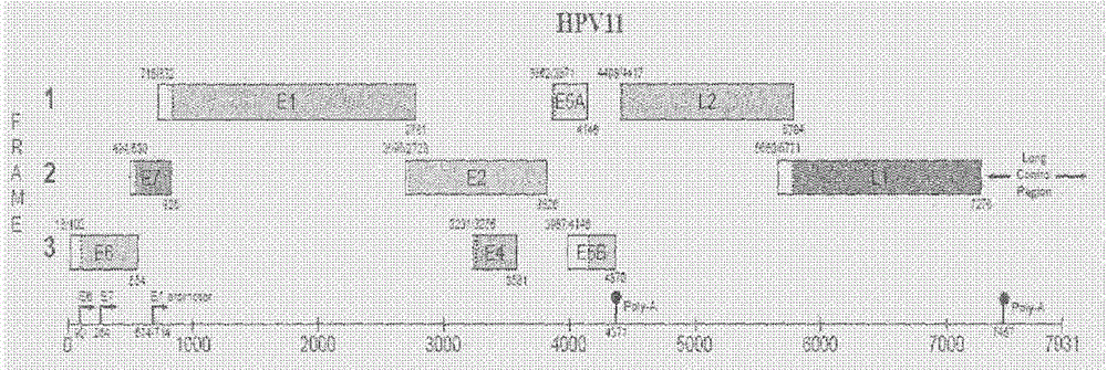
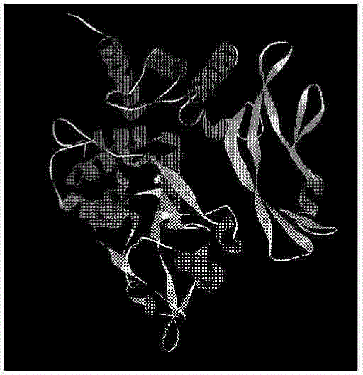
[0038] Using the Protein Data Bank (PDB) database, the three-dimensional crystal structure of the HPV18 E1 protein required for our experiment ( image 3 ) to download and get the amino acid sequence of E1 protein (FASTA format, figure 2 ), so as to carry out the next operation.

[0039] 2. Use Autodock molecular docking software to determine the active center and set the active pocket according to the active tyrosine residue site Y492 of E1

[0040] In this step, firstly, a known molecule that can undergo an addition reaction with the active tyrosine residue site Y492 of E1 is used as a positive control molecule. The binding energy of the interaction and t...

Embodiment 2

[0076] Example 2 A virtual screening of anti-HPV virus drugs by using R589 of the E1 protein as the active site to construct an active pocket.

[0077] The method is the same as in Example 1, the difference is: when setting the active pocket: the central coordinate of R589 is the center of the active pocket, and finally the conditions for setting the active pocket are as follows: the active tyrosine residue site R589 of E1 is used as the active pocket. Active pocket in the center: the center coordinates are 36.410 for x, 25.423 for y, and 106.056 for z, the grid size is 40×40×40, and the spacing is 0.375nm.


PUM


Abstract

The invention relates to an anti-HPV (human papillomavirus) medicine virtual screening method by using DNA (deoxyribonucleic acid) helicase E1 of HPV as a target point. The method comprises the following steps that 1) PDB (protein data bank) structure data of an E1 protein structure is determined; 2) molecular docking software is adopted, the active center is determined according to active amino acid residue sites of E1, and active bags are set; 3) micromolecular ligands are screened; 4) a micromolecular ligand database for docking is built; 5) the micromolecular ligands in the micromolecular ligand database for docking are in one-to-one docking with the active bags by the molecular docking software according to the set active bags; and 6) pilot medicine with the cervical cancer resistance effect is preliminarily determined according to the docking result sequencing. When the anti-HPV medicine virtual screening method is adopted, a clue of active compounds can be obtained in the short time, then, the cervical cancer resistance medicine is screened by the animal level or a high-flux cell platform, the speed and the efficiency are greatly accelerated and improved, and the development period of new medicine is shortened.

Description

technical field [0001] The invention relates to a drug screening method, in particular to a virtual screening method for novel anti-HPV virus drugs using molecular docking with HPV E1 as the target. Background technique [0002] Cervical cancer is a disease that seriously endangers women's health and life. There are about 500,000 new cases and 250,000 deaths every year. It is the second leading cause of women's death, most of which are in developing countries, accounting for about 80%. Cervical cancer is a type of tumor with the highest incidence rate among malignant tumors of female reproductive tract in my country. According to statistics, there are about 135,000 new cases of cervical cancer in my country every year, and about 50,000 of them die as a result, which seriously threatens the physical and mental health and life safety of women. In 1977, German scholar Zur Hausen et al. discovered human papillomavirus (human papillomavirus, HPV) DNA from cervical cancer specime...

Claims


Application Information

Patent Timeline
IPC IPC(8): G06F19/16A61K45/00A61P35/00A61P31/20
Inventor 芦秀丽高兵张勇陈树超刘汀贾丹
Owner 南京益添生物科技有限公司
Learn More