A preparation method of esophageal cancer immunomass spectrometry detection kit
A detection kit and mass spectrometry detection technology, which is applied in the field of biotechnology and mass spectrometry detection, can solve problems such as not being suitable for population censuses, and achieve the effects of strong binding specificity, simple operation, and low cost
- Summary
- Abstract
- Description
- Claims
- Application Information
AI Technical Summary
Problems solved by technology
Method used
Image
Examples
Embodiment 1
[0036] Example 1 Preparation method of polypeptide marker antigen monoclonal antibody
[0037] 1) BALB / C mice were immunized with synthetic polypeptide 5 coupled to carrier protein KLH as an immunogen; wherein, the full-length sequence of polypeptide 5 was: Asn-Leu-Gly-His-Gly-His-Lys-His-Glu -Arg-Asp-Gln-Gly-His-Gly-His-Gln.
[0038]2) After 2 weeks, the tail blood titer was detected, and when it reached above 1:1000, splenocytes from BALB / C mice were fused with SP2 / 0 mouse myeloma cells under the action of PEG.
[0039] 3) Screen by ELISA method, clone and purify hybridoma cells positive for secreting antibodies by limited dilution method.
[0040] 4) Screen out 10 hybridoma cells against synthetic peptide 5, one of which is highly sensitive (1ng / well), expand the cell line, prepare and purify monoclonal antibody ascites, and test the titer of monoclonal antibody (1:200000 ), titer (0.0005μg / ml) and subtype identification (IgG2b), etc., to obtain specific monoclonal antibo...
Embodiment 2
[0041] Example 2 Esophageal cancer immune mass spectrometry detection method
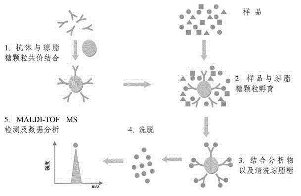
[0042] The flow process of the polypeptide immune mass spectrometry detection method of the present invention is as follows: figure 1 shown. Specifically:
[0043] 1) Take 25 μL protein G-coated agarose particles (Protein G Agarose, Santa Cruz), put it in a 0.6 mL Eppendorf tube, add 7.78 μg AP0105, rotate at 4°C (speed 5r / min), mix for 1 hour, and let stand for 3 minutes , remove the supernatant, and wash Protein G Agarose 3 times with 100 μL PBS buffer (0.01mol / l, pH7.4);
[0044] 2) Take 24 μg of the peptide standard product of synthetic peptide 5 (solid-phase chemical synthesis, Beijing Zhongke Yaguang Biotechnology Co., Ltd.), 50 μL of PBS, mix with the Protein G Agarose washed in step 1), and rotate and mix at 4°C for 8 hours;
[0045] 3) Leave it for 3 minutes, remove the supernatant, and wash Protein G Agarose 3 times with 200 μL PBS; transfer the suspension to another clean Eppendorf tu...
Embodiment 3
[0050] Example 3 Esophageal cancer immune mass spectrometry detection method
[0051] The flow process of the polypeptide immune mass spectrometry detection method of the present invention is as follows: figure 1 shown. Specifically:
[0052] 1) Take 15 μL protein A-coated agarose particles (Protein A Agarose, Santa Cruz), put it in a 0.2 mL Eppendorf tube, add 1.56 μg AP0105, rotate at 4°C (speed 5r / min), mix for 15 minutes, and place for 2 minutes. Remove the supernatant and wash Protein A Agarose 3 times with 100 μL PBS buffer (0.01mol / l, pH7.4);
[0053] 2) Take 24 μg of synthetic peptide 5 polypeptide standard (solid-phase chemical synthesis, Beijing Zhongke Yaguang Biotechnology Co., Ltd.), 50 μL of PBS, mix with the Protein A Agarose washed in step 1), and mix at 4°C for 8 hours;
[0054] 3) Leave it for 2 minutes, remove the supernatant, and wash Protein A Agarose 3 times with 200 μL PBS; transfer the suspension to another clean Eppendorf tube for the last wash;
...
PUM
| Property | Measurement | Unit |
|---|---|---|
| Sensitivity | aaaaa | aaaaa |
Abstract
Description
Claims
Application Information
 - R&D
- Intellectual Property
- Life Sciences
- Materials
- Tech Scout
- Unparalleled Data Quality
- Higher Quality Content
- 60% Fewer Hallucinations
Browse by: Latest US Patents, China's latest patents, Technical Efficacy Thesaurus, Application Domain, Technology Topic, Popular Technical Reports.
© 2025 PatSnap. All rights reserved.Legal|Privacy policy|Modern Slavery Act Transparency Statement|Sitemap|About US| Contact US: help@patsnap.com


