Hydrogel-nanometer fiber membrane, preparation method and uses thereof
A nanofiber membrane and nanofiber technology, applied in the field of biomedicine, to achieve sufficient permeability, convenient surgical operation, and good permeability
- Summary
- Abstract
- Description
- Claims
- Application Information
AI Technical Summary
Problems solved by technology
Method used
Image
Examples
Embodiment 1
[0057] 1) Respectively mix 200μL chondroitin sulfate prepared above, 25μL H 2 O 2 Add 25μL horseradish peroxidase (HRP) to a 1.5mL centrifuge tube and mix well;
[0058] 2) Add the mixture prepared in step (1) to a 24-well cell culture plate, add 100μL to each well, at 37°C, 5% CO 2 Place in the incubator for 4 hours; add 0.5 mL of medium to each hole and put it in the incubator overnight;
[0059] 3) Aspirate the medium of the cell culture plate in step (2), spread 0.5mL Matrigel per well to facilitate the growth of RPE cells, put it in the incubator for 2 hours, and set aside;
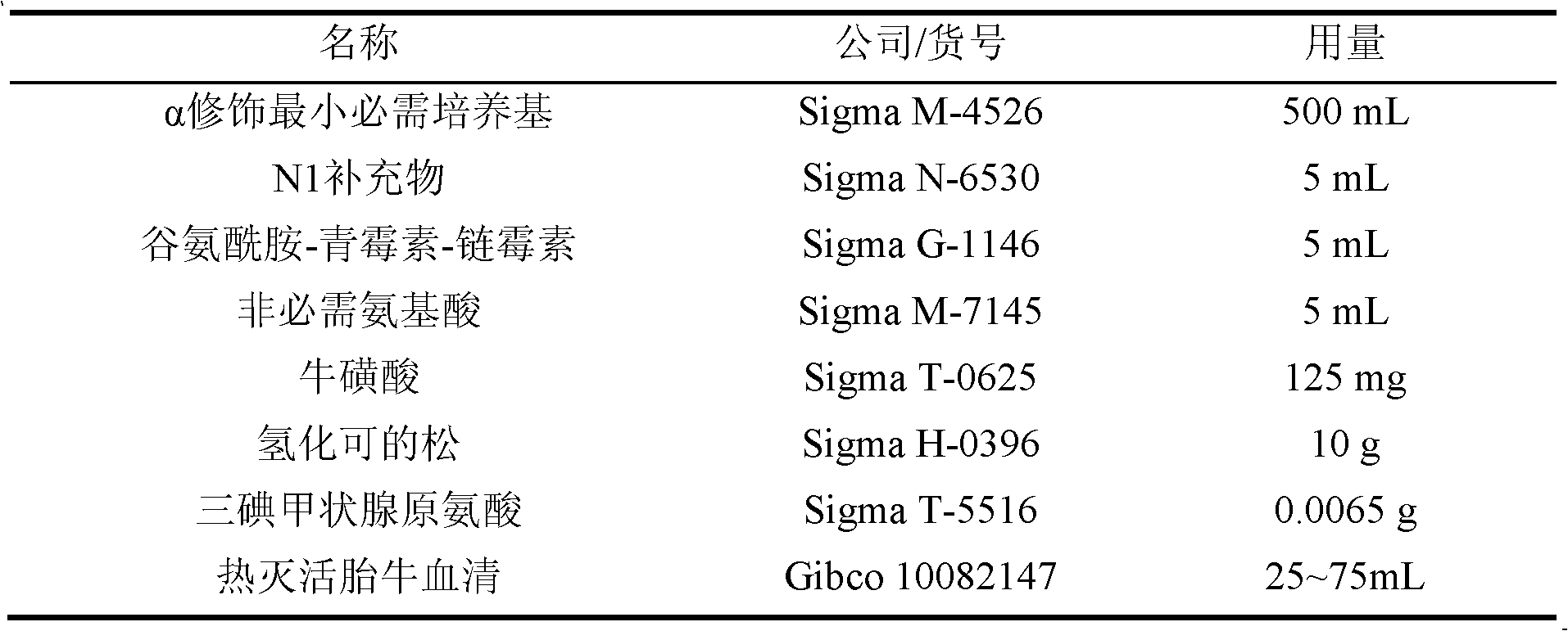
[0060] 4) Aspirate the culture medium in the cultured RPE cell culture plate (calculated as 6-well cell culture plate), add 1mL PBS to each well to remove the remaining medium, after aspirating PBS, add 0.5mL per well Put 0.25% pancreatin-EDTA into the incubator for 2-5 minutes. After 85-95% of the RPE cells are detached from the plate wall, add 1 mL of medium to each well to stop the pancreatin-EDTA effect...
Embodiment 2
[0064] 1) Respectively mix 200μL chondroitin sulfate prepared above, 25μL H 2 O 2 Add 25μL horseradish peroxidase (HRP) to a 1.5mL centrifuge tube and mix well;
[0065] 2) Add the mixture prepared in step (1) to a 24-well cell culture plate, add 100μL to each well, at 37°C, 5% CO 2 Place it in the incubator for 3 hours; add 1 mL of medium to each hole and put it in the incubator overnight;
[0066] 3) Aspirate the culture medium of the cell culture plate in step (2), spread 1mL Matrigel per well to facilitate the growth of RPE cells, and put it in the incubator for 3 hours for use;
[0067] 4) Aspirate the culture medium in the RPE cell culture plate (calculated as the 6-well cell culture plate), add 0.5mL PBS to each well to remove the remaining medium, after aspirating the PBS, add 0.75mL per well It is 0.25% pancreatin-EDTA, put it in the incubator for 2-5 minutes, after 85-95% RPE cells are detached from the plate wall, add 1.5mL medium to each well to stop the pancreatin-EDTA e...
Embodiment 3
[0071] 1) Respectively mix 200μL chondroitin sulfate prepared above, 25μL H 2 O 2 Add 25μL horseradish peroxidase (HRP) to a 1.5mL centrifuge tube and mix well;
[0072] 2) Add the mixture prepared in step (1) to a 24-well cell culture plate, add 100μL to each well, at 37°C, 5% CO 2 Place it in the incubator for 5 hours; add 2 mL of medium to each hole and put it in the incubator overnight;
[0073] 3) Aspirate the culture medium of the cell culture plate in step (2), spread 2mL Matrigel per well to facilitate the growth of RPE cells, and put it in the incubator for 4 hours for use;
[0074] 4) Aspirate the culture medium in the RPE cell culture plate (calculated as the 6-well cell culture plate), add 2mL PBS to each well to remove the remaining medium, after aspirating the PBS, add 1mL per well to the content of 0.25 % Trypsin-EDTA, put it in the incubator for 2-5 minutes, after 85-95% of RPE cells are detached from the plate wall, add 2 mL of medium per well to stop the effect of t...
PUM
| Property | Measurement | Unit |
|---|---|---|
| thickness | aaaaa | aaaaa |
Abstract
Description
Claims
Application Information
 - R&D
- Intellectual Property
- Life Sciences
- Materials
- Tech Scout
- Unparalleled Data Quality
- Higher Quality Content
- 60% Fewer Hallucinations
Browse by: Latest US Patents, China's latest patents, Technical Efficacy Thesaurus, Application Domain, Technology Topic, Popular Technical Reports.
© 2025 PatSnap. All rights reserved.Legal|Privacy policy|Modern Slavery Act Transparency Statement|Sitemap|About US| Contact US: help@patsnap.com

