
Method for inducing synovium mesenchymal stem cells to be differentiated to chondrocytes by in-vitro lentivirus mediated BMP-2 (Bone Morphogenetic Protein) genes

A BMP-2, mesenchymal stem cell technology, applied in the field of biomedicine, can solve the problems of high time and cost, low success rate, etc.

Inactive Publication Date: 2013-06-12
SECOND MILITARY MEDICAL UNIV OF THE PEOPLES LIBERATION ARMY
View PDF0 Cites 6 Cited by
  • Summary
  • Abstract
  • Description
  • Claims
  • Application Information

AI Technical Summary

Problems solved by technology

[0005] The purpose of the present invention is to provide a method for in vitro lentivirus-mediated BMP-2 gene-induced synovial mesenchymal stem cells to differentiate into chondrocytes, said in vitro lentivirus-mediated BMP-2 gene-induced synovial mesenchymal stem cells The method for differentiation of mesenchymal stem cells to chondrocytes needs to solve the technical problems of low success rate, high time and cost in the prior art of inducing synovial mesenchymal stem cells to differentiate into chondrocytes

Method used


Image

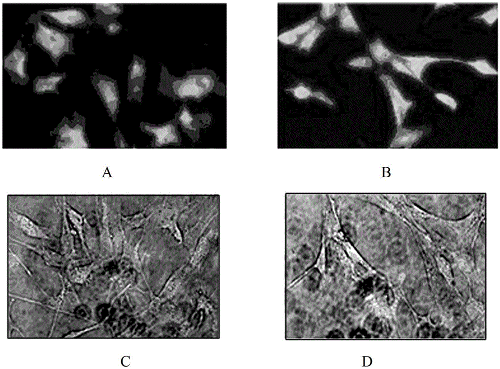
  • Method for inducing synovium mesenchymal stem cells to be differentiated to chondrocytes by in-vitro lentivirus mediated BMP-2 (Bone Morphogenetic Protein) genes
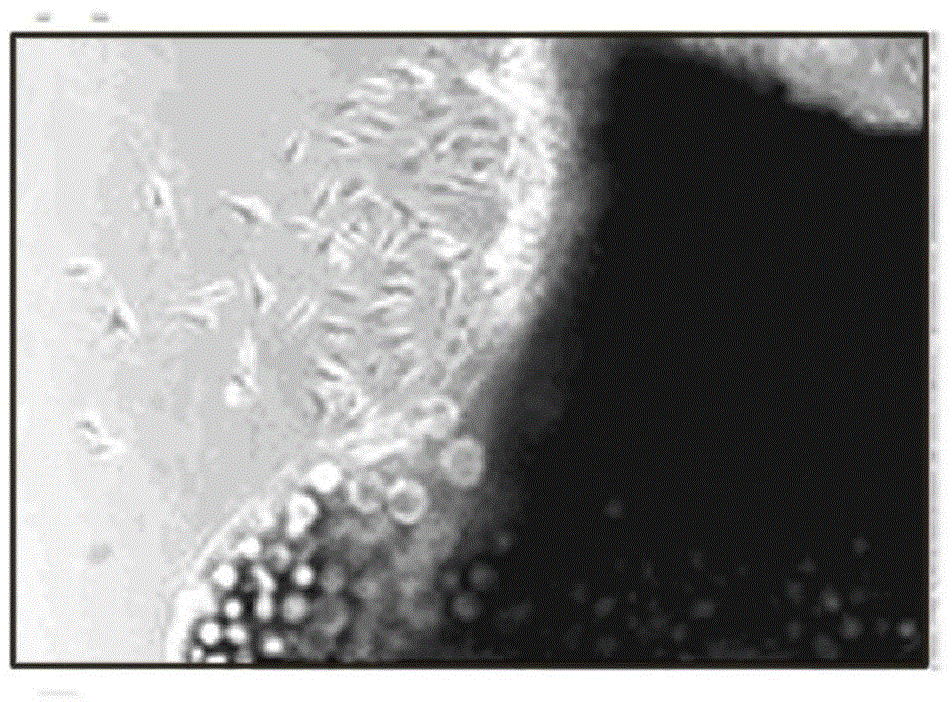
  • Method for inducing synovium mesenchymal stem cells to be differentiated to chondrocytes by in-vitro lentivirus mediated BMP-2 (Bone Morphogenetic Protein) genes
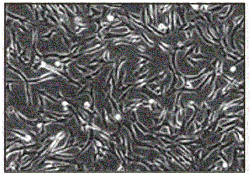
  • Method for inducing synovium mesenchymal stem cells to be differentiated to chondrocytes by in-vitro lentivirus mediated BMP-2 (Bone Morphogenetic Protein) genes

Examples


Embodiment 1

[0031] Embodiment 1: the acquisition, separation and identification of SMSCs

[0032] 1.1 Get SMSCs

[0033] Six 3-month-old New Zealand white rabbits (SPF grade, Slack Experimental Animal Center, Chinese Academy of Sciences, http: / / www.slaccas.com ) (average weight 2.1±0.3kg, male or female). After fixed anesthesia, a straight parapatellar incision was taken, and the joint capsule was opened layer by layer, and the suprapatellar bursa synovial tissue was harvested with tissue scissors. The obtained synovium was cut into pieces, and collagenase type 2 (0.2%, Sigma-Aldrich, Inc., http: / / www.sigmaaldrich.com ) digested for 3 hours, passed through a 200-lm filter (Jiabao Company, http: / / www.caigou.com.cn / c20931 ) to obtain tissue pieces after filtration, and cultured in 20% FBS (Hyclone company http: / / www.hyclone.com ) DMEM (GibcolBRL, http: / / www.gelcompany.com / products / gibco-brl )middle. All the above processes were carried out in the ultra-clean bench (SW-CJ-1F, Jiab...

Embodiment 2

[0052] Example 2 Vector construction and transfection

[0053] 2.1 Primer design

[0054] The GenBank number of rabbit BMP-2 is NM_0010826507, and the reading frame is 70-1257bp. Accordingly, using the SGD software ( http: / / www.yeastgenome.org / cgi-bin / web-primer ) to design primers. The primer was designed at the 5' end: 5'-CTAGCTAGCTGGAGGCTCTTTCAATGG-3' (SEQ ID NO: 1). GCTAGC is the enzyme cutting site of the restriction endonuclease Nhe1, ATG is the stop codon; 3' end:

[0055] 5'-GGGTTTACCGGTAAAGCAAAAACAAACCAA-3' (SEQ ID NO: 2). ACCGGT is the cutting site of restriction endonuclease Age1, and AAG is the stop codon.

[0056] 2.2 Amplification of BMP-2

[0057] Trizol (Gibco, http: / / zh.invitrogen.com ) to extract total RNA from rabbit liver, using 5IU MMLV (karma company, http: / / www.karmabio.cn) for the reverse transcription reaction. cDNA was synthesized after a cycle of reaction at 70°C for 5 minutes, 37°C for 60 minutes, and 95°C for 5 minutes. oBMP-2 was ampl...

Embodiment 3

[0068] SMSCs security identification after embodiment 3 transfection

[0069] 3.1 Analysis of cell proliferation kinetics

[0070] The transfected SMSCs were pressed at 50cells / cm 2 After planting in a 24-well plate, put CO 2 in the incubator. Select 3 wells every day, digest with 200 μL of 0.25% Trypsin / EDTA, and pipette repeatedly to form a single-cell suspension. The medium was not changed until all wells were taken. The number of cells in each well was counted every day and recorded as the mean value. According to the value, the growth curve is drawn according to the equation and the doubling time is calculated in turn. The equation used is TD=tlog2 / log(N / N0) (t: the time from the beginning of culture to the counted day, in hours; N: the cell count at time T; N0: the total number of cells).

[0071] Results: The number of cells for 7 days was counted, and the cell growth curve was drawn accordingly. According to the relevant formula, the cell doubling time was calcu...


PUM


Abstract

The invention relates to a method for inducing synovium mesenchymal stem cells (SMSCs) to be differentiated to chondrocytes by in-vitro lentivirus mediated BMP-2 (Bone Morphogenetic Protein) genes. The method comprises the following steps of: separating and culturing synovium mesenchymal stem cells; constructing a recombinant plasmid pFUGW-oBMP-2; preparing morbus virosus of transfected lentivirus; and transfecting synovium mesenchymal stem cells by the morbus virosus of transfected lentivirus, wherein in the step of preparing morbus virosus of transfected lentivirus, the transfected lentivirus is jointly formed by the recombinant plasmid pFUGW-oBMP-2 and a packaging plasmid; and in the step of transfecting synovium mesenchymal stem cells by the morbus virosus of transfected lentivirus, synovium mesenchymal stem cells over third generation is taken to be mixed with the morbus virosus of transfected lentivirus, and then added into incomplete chondroblast inducing culture liquid to induce so as to obtain chondrocytes. SMSCs transfected by the transfected lentivirus provided by the invention are safe enough and can be spontaneously differentiated to cartilage in vitro.

Description

Technical field: [0001] The present invention relates to the field of biomedicine, in particular to a method for cartilage differentiation, in particular to a method for in vitro lentivirus-mediated BMP-2 gene-induced synovial mesenchymal stem cells to differentiate into chondrocytes. Background technique: [0002] In recent years, meniscal cartilage repair using synovial mesenchymal stem cells has become a hot topic in joint surgery. The classic tissue engineering method is to induce stem cells to proliferate in vitro to form seed cells, plant the seed cells on the biological scaffold, move the scaffold back to the organism and add various cytokines for repair. [0003] In the past, many researchers chose bone marrow-dervied mesenchymal stem cells (BMSCs) as seed cells. But when De Barii discovered synovium-dervied mesenchymal stem cells (SMSCs), they quickly replaced BMSCs. The reason is that although SMSCs are similar to BMSCs in terms of cell morphology, immunophenotyp...

Claims


Application Information

Patent Timeline
IPC IPC(8): C12N15/867C12N5/10
Inventor 符培亮张雷丁喆如吴海山吴宇黎丛锐军陈松
Owner SECOND MILITARY MEDICAL UNIV OF THE PEOPLES LIBERATION ARMY
Learn More