Early stage cervical carcinoma detection system integrating fluorescent mesoscope imaging and optical coherence tomography (OCT)

A detection system and imaging system technology, applied in the field of biomedical engineering, can solve the problems that cannot meet the requirements of CIN2 or CIN3 diagnosis, and achieve the effects of improving diagnostic specificity, deep detection depth, and reducing morbidity

Inactive Publication Date: 2013-06-19
TIANJIN UNIV
View PDF6 Cites 32 Cited by
  • Summary
  • Abstract
  • Description
  • Claims
  • Application Information

AI Technical Summary

Problems solved by technology

In terms of optical imaging of cervical cancer in vivo, Peking University Shenzhen School of Medicine has studied the improvement of sensitivity of OCT imaging-assisted acetowhite diagnosis. Imaging methods are not yet adequate for the diagnosis of CIN2 or CIN3

Method used


Image

  • Early stage cervical carcinoma detection system integrating fluorescent mesoscope imaging and optical coherence tomography (OCT)
  • Early stage cervical carcinoma detection system integrating fluorescent mesoscope imaging and optical coherence tomography (OCT)
  • Early stage cervical carcinoma detection system integrating fluorescent mesoscope imaging and optical coherence tomography (OCT)

Examples


Embodiment Construction

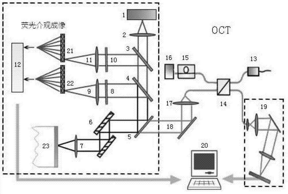
[0037] The invention provides a combined fluorescence mesoscopic imaging and OCT imaging system for early cervical cancer detection. On the basis of high-sensitivity imaging of the whole cervix by colposcopy, local high-spatial-resolution morphological imaging and medium-spatial High-resolution functional imaging to improve the specificity of cervical cancer diagnosis with complementary information fused with function and structure.

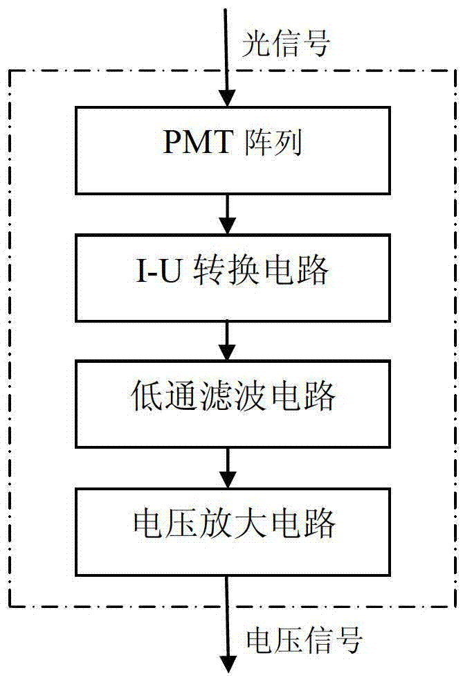
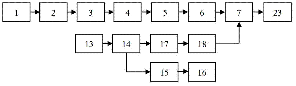
[0038] see figure 1 The fluorescent mesoscopic imaging and OCT combined imaging system suitable for early cervical cancer detection proposed by the present invention includes ultraviolet laser source and transmission, turning, optical path for changing laser polarization, detector for positioning receiving beam and detector control processing Part, the fluorescence mesoscopic imaging system of the data acquisition card; including the spectral OCT system including the near-infrared laser source, fiber optic beam splitter and spectrometer; includin...


PUM


Abstract

The invention belongs to the technical field of biomedical engineering, and relates to an integration imaging system used for early stage cervical carcinoma detection and integrating fluorescent mesoscope imaging and optical coherence tomography (OCT). The integration imaging system comprises a fluorescent mesoscope imaging system and a spectrum OCT system. The fluorescent mesoscope imaging system comprises a laser light source, a polarizer, a polarized light spectroscope, a first dichroscope, a second dichroscope, an X-Y scanning galvanometer, an objective lens, a fluorescent detection part, a diffused light detection part and a computer, wherein the X-Y scanning galvanometer and the objective lens are shared by the spectrum OCT system. The spectrum OCT system comprises a low-coherent light source, a fiber polarization splitter, a focusing lens, the X-Y scanning galvanometer, the objective lens and a spectrograph. Ultraviolet light beams generated by the laser light source pass through the polarizer, the polarized light spectroscope and the first dichroscope and then are reflected through the second dichroscope, and samples generated by the low-coherent light source are collimated through the focusing lens and are combined with the ultraviolet light beams. The system improves diagnostic specificity through mutual evidence of organization function information and histomorphology information.

Description

technical field [0001] The invention belongs to the technical field of biomedical engineering and relates to an early cervical cancer detection system. Background technique [0002] Cervical cancer is the second most common malignant tumor in women. Every year, there are about 510,000 new cases of cervical cancer in the world, and about 300,000 women die of cervical cancer. One of the main pathogenic factors of cervical cancer is HPV infection, a small number of HPV infection will develop into cervical intraepithelial neoplasia 1 (CIN1), 10-20% of CIN1 will develop into more serious CIN2, CIN3, 5% -12% of CIN2 and CIN3 will develop into cancer within 8-10 years. Once invasive cancer is formed, it will grow rapidly. If not diagnosed and treated in time, the patient will die within 2-5 years. Therefore, it is very necessary to carry out correct diagnosis and treatment in CIN2 and CIN3 stages. [0003] The current clinical method for diagnosing cervical cancer is: first use P...

Claims


Application Information

Patent Timeline
Patent Type & Authority Applications(China)
IPC IPC(8): G01N21/64G01N21/19
Inventor 赵会娟崔姗姗贾梦宇高峰
Owner TIANJIN UNIV
Why Eureka
Try Eureka