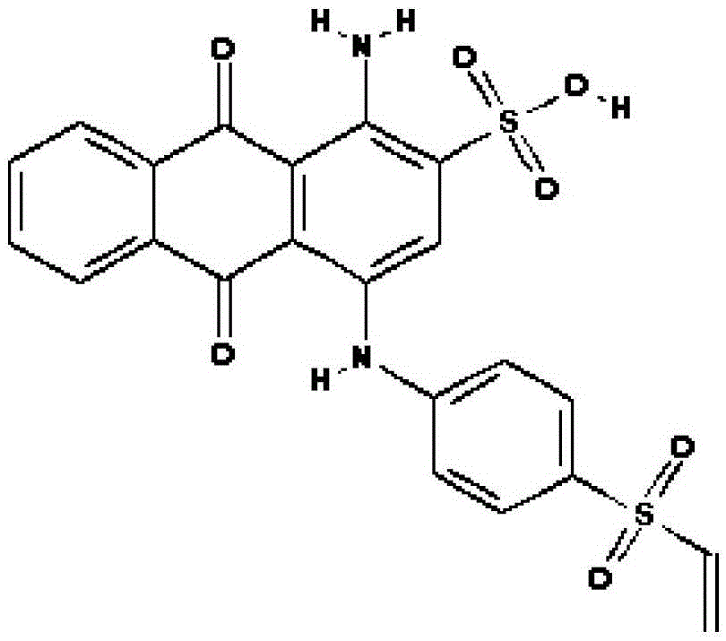
The use of single blue a as a protein prestain
A pre-staining agent and protein technology, which is applied in the field of single blue A as a protein pre-staining agent, can solve the problems of high cost and many detection steps, and achieve the effect of moderate sensitivity, simple operation and low price
- Summary
- Abstract
- Description
- Claims
- Application Information
AI Technical Summary
Problems solved by technology
Method used
Image
Examples
Embodiment 1
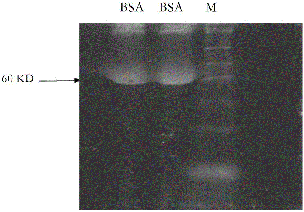
[0096] Example 1 Various staining methods and protein purification after staining with single blue A staining protein
[0097] 1. Method of staining protein with single blue A
[0098] 1.1 Heating catalyzed dyeing reaction
[0099] (1) Mix 10 μl single blue A staining solution with 90 μl protein solution.
[0100] (2) Heat the mixed sample at 100°C for 1 min.
[0101] (3) Then add 100 μl of dyeing optimizer and heat at 100°C for 1 min.
[0102] (4) After the sample is cooled to room temperature, add 20 μl staining terminator and let stand at room temperature for 5 minutes.
[0103] 1.2 Electrical stimulation catalyzed staining reaction
[0104] (1) Take 10 μl single blue A staining solution and 90 μl 10% BSA protein solution and mix thoroughly.
[0105] (2) Add the mixed sample to a 200ml electro-cup.
[0106] (3) The electrorotator selects 1800V as the output voltage and shocks for 20ms.
[0107] (4) Then add 100 μl dyeing optimizer and let stand at room temperature fo...
experiment example 1
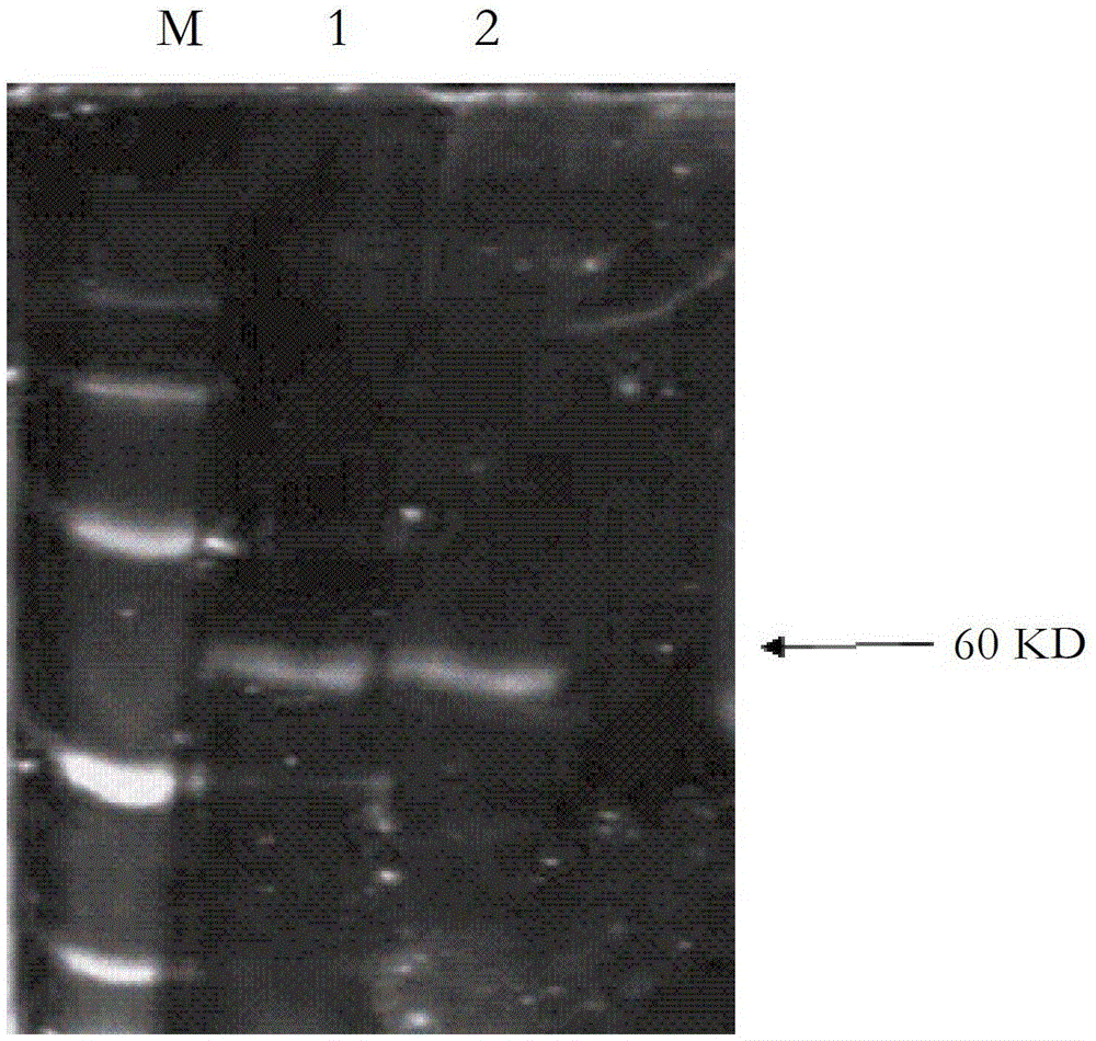
[0128] Experimental example 1 Preparation and identification of prestained protein markers with single blue A dye
[0129] 1 Experimental method
[0130] 1.1 marker protein mix
[0131] Add 0.1 μg of Taq enzyme protein, BSA protein, lactate dehydrogenase A protein and IL-7 into 1.5ml EP tubes and mix thoroughly to obtain unstained mixed protein molecular weight standard protein (marker protein).
[0132] 1.2 Marker protein staining
[0133] (1) Add 3 μl of Mono Blue A dye and mix thoroughly.
[0134] (2) Heat the mixed sample at 100°C for 1 min.
[0135] (3) Then add 30 μl of dyeing optimizer and heat at 100°C for 1 min.
[0136] (4) After the sample is cooled to room temperature, add 0.6 μl staining terminator and let stand at room temperature for 5 minutes.
[0137] 1.3marker protein purification
[0138] (1) Weigh 5g of Dextran G-50, dilute to 50ml with deionized water, boil in boiling water for 3h.
[0139] (2) Put the well-stirred gel into a 0.5ml EP tube with a ho...
experiment example 2
[0153] Experimental example 2 Gradient detection experiment of single blue A labeled bovine serum albumin
[0154] 1. Experimental method
[0155] Take three 1.5ml EP tubes respectively, add 10 μl, 1 μl, 0.1 μl of BSA protein with a concentration of 1 μg / μl, and then add 9 μl of double-distilled water to the EP tube with 1 μl of BSA protein at a concentration of 1 μg / μl, and add 0.1 Add 9.9 μl of double-distilled water to the EP tube of BSA protein with a concentration of 1 μg / μl, and mix thoroughly. Take 1 μl of the solution from each tube, and add 1 μl of monoblue A dye (200mM) respectively, and heat at 100°C for 1min. After the samples were cooled, 10 μl of dyeing optimizer was added to each tube, and heated at 100°C for 1 min. After the 3 tubes were cooled to room temperature, 2 μl of staining terminator was added respectively, and the tubes were allowed to stand at room temperature for 5 minutes. Subsequent identification by SDS-PAGE electrophoresis.
[0156] 2. Exper...
PUM
 Abstract
Description
Claims
Application Information
 - R&D
- Intellectual Property
- Life Sciences
- Materials
- Tech Scout
- Unparalleled Data Quality
- Higher Quality Content
- 60% Fewer Hallucinations
Browse by: Latest US Patents, China's latest patents, Technical Efficacy Thesaurus, Application Domain, Technology Topic, Popular Technical Reports.
© 2025 PatSnap. All rights reserved.Legal|Privacy policy|Modern Slavery Act Transparency Statement|Sitemap|About US| Contact US: help@patsnap.com


