Supercharge Your Innovation With Domain-Expert AI Agents!

Method for detecting cells from a sample

A cell and sample technology, applied in the field of detecting cells from samples, can solve problems such as the importance not mentioned

Inactive Publication Date: 2016-06-08
FZMB医疗技术和生物技术研究中心有限公司
View PDF11 Cites 0 Cited by
  • Summary
  • Abstract
  • Description
  • Claims
  • Application Information

AI Technical Summary

Problems solved by technology

The reference does not mention the importance of penetration of the components to be analyzed into the pores of the porous solid phase

Method used


Image

  • Method for detecting cells from a sample
  • Method for detecting cells from a sample
  • Method for detecting cells from a sample

Examples


Embodiment 1

[0068] Legionella in drinking water ( Legionella ) for fluorescence detection.

[0069] 1. Functionalization of PTFE membrane and antibody immobilization.

[0070] In the first step, PTFE membranes hydrophilized by OH groups (OMNIPOREJC from Millipore) were aminated by treatment with 3-aminopropyltriethoxysilane (APTS). Therefore, 50 μl of 3-aminopropyltriethoxysilane (Sigma-Aldrich Chemie GmbH) was dissolved in 4950 μl of acetone and applied to the dried membrane (d=5 cm) in a 90 mm glass Petri dish. The dishes were transferred to a glass desiccator which was rapidly (<10 min) evacuated to <300 mbar. Incubate for 12 hours at room temperature at 20 rpm on an orbital shaker in a closed desiccator. Subsequently, 0.42 μl of reagent grade 37% hydrochloric acid (Carl Roth GmbH & Co. KG) was added. The dishes were then transferred to a glass desiccator which was rapidly (<10 min) evacuated to <300 mbar. Then incubate for 6 hours at room temperature at 20 rpm on an orbital shak...

Embodiment 2

[0082] Via HRP / TMB prec. Drinking water tested Legionella Detection of bacteria.

[0083] 1. Antibody immobilization on HDPE membrane.

[0084] In a first step, anti- Legionella Antibodies (5F4, Senova, Weimar) were immobilized by physisorption on HDPE membranes (Porex Inc. type T3, pore size 5-10 μm, thickness 200 μm, high density polyethylene = HDPE). Therefore, 50 round filters (d=5 mm) were punched out and placed in a 50 ml beaker with 20 ml ethanol (96%) and degassed in a desiccator at 10 Torr, on a magnetic stirrer Stir gently for 20 min. Subsequently, the filter was washed with 50% ethanol solution and finally degassed three times in 10 ml of carbonate buffer (100 mM, pH 9.5). Subsequently, 100 μl of 1 mg / ml anti- Legionella solution. The filters were incubated in a desiccator for 6 hours at room temperature with gentle agitation. Subsequently, the antibody solution was replaced with a block solution (5.5 mg / ml casein dissolved in PBS buffer (150 mM, pH=7.3))...

Embodiment 3

[0090] by vital staining on a hyaline membrane Salmonella Bacteria detection.

[0091] 1. Immobilization of antibodies.

[0092] The membrane according to item 1 of example 1 was used. Instead of the antibody used therein, monoclonal anti- Salmonella Bacteria antibody (1E6, SifinGmbH, Berlin) was immobilized on the membrane.

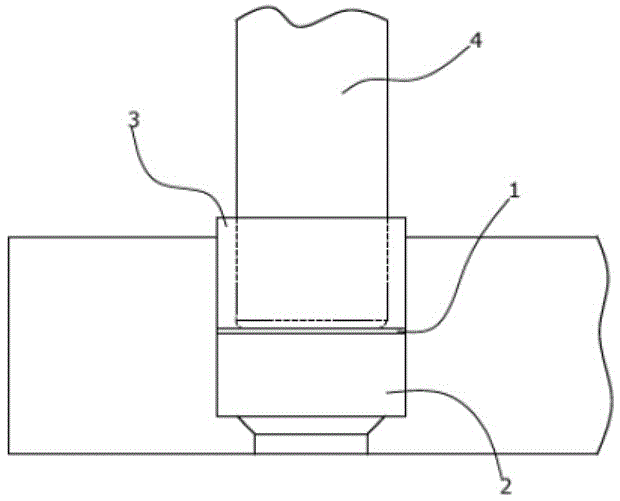
[0093] 2. Preparation of the filter assembly.

[0094] A circular filter with a diameter of 24 mm was punched out of the membrane using a hole punch. The filter was fixed to the bottom of an empty 20 ml column with an insertable bottom (SpinColumn, AHNBiotechnology, Nordhausen, Germany). A peristaltic pump was connected to the lower luer port of the column.

[0095] 3. Sample preparation.

[0096] 25g of heat-sterilized meat samples and 225ml of buffered peptone water were added to the inner filter bag of a Stomacherbag (BA6141 / STR, SewardLimited, UK), and 10 μl of Salmonella typhimuria (ATCC14028) pre-culture (1000germs / ml) incubation. The sam...


PUM

PropertyMeasurementUnit
thicknessaaaaaaaaaa
lengthaaaaaaaaaa
widthaaaaaaaaaa

Abstract

A method for detecting cells from a sample comprising the steps of: (a) applying a sample or portion of a sample in a liquid phase to a porous, generally two-dimensional support having pores in the direction of flow having on its surface at least one binding molecule specific for the cells to be detected or fragments of these cells; (b) enabling the cells to be detected or fragments of these cells to enter the pores of said porous generally two-dimensional carrier from the sample or part of the sample wherein, the average pore size of the pores is such that the cells or fragments to be detected can enter the pores; (c) the cells or fragments to be detected are retained by at least one binding molecule specific to the cells or fragments to be detected; (d) the cells or fragments to be detected are retained by The imaging method performs an optical readout method on said generally two-dimensional support, optionally after the cells or cell fragments to be detected have been labeled with a labeling agent; wherein (e) used for the readout method includes, for example, a lens The optical axis of the optical system of the system generally extends in the direction of flow of the sample or sample part; and (f) detection of cells or cell fragments is performed on the surface of said porous generally two-dimensional support.

Description

technical field [0001] The present invention relates to a method for detecting cells, in particular microorganisms, from a sample. Background technique [0002] Quantitative and qualitative detection of cells in complex sample solutions is a fundamental operation in microbiology, cytology and modern biotechnology. [0003] In this respect, the rapid and direct detection of microorganisms in food samples is particularly stringent. Due to the potentially high complexity of the sample matrix, but also bacteria of particular concern (e.g. Salmonella ( Salmonella ), Listeria ( Listeria ) or Legionella ( Legionella ) (drinking water)) in terms of the need to ensure sterility, and therefore the detection limit of bacteria, it has high requirements. In particular, the food industry requires a process that does not require a step of enrichment, ie the propagation of microorganisms. It is a problem of great practical significance to provide new rapid and cheap microbial detecti...

Claims


Application Information

Patent Timeline
Patent Type & Authority Patents(China)
IPC IPC(8): G01N33/543G01N33/569
CPCG01N33/54313G01N33/5436G01N33/54366G01N33/54386G01N33/569G01N33/56911
Inventor 斯蒂芬·亨策彼得·米尔希
Owner FZMB医疗技术和生物技术研究中心有限公司
Features
  • R&D
  • Intellectual Property
  • Life Sciences
  • Materials
  • Tech Scout
  • Unparalleled Data Quality
  • Higher Quality Content
  • 60% Fewer Hallucinations
Learn More