Strong signal and low toxicity composite nanometer material and preparation method thereof

A nanomaterial and composite technology, applied in nanotechnology, nanotechnology, nano-optics, etc. for materials and surface science, to achieve the effect of low toxicity

Inactive Publication Date: 2014-03-19
CHANGCHUN UNIV OF SCI & TECH
View PDF2 Cites 9 Cited by
  • Summary
  • Abstract
  • Description
  • Claims
  • Application Information

AI Technical Summary

Problems solved by technology

This invention has

Method used


Image

  • Strong signal and low toxicity composite nanometer material and preparation method thereof
  • Strong signal and low toxicity composite nanometer material and preparation method thereof
  • Strong signal and low toxicity composite nanometer material and preparation method thereof

Examples


Embodiment 1

[0028] Embodiment 1 Preparation of a composite nanoparticle:

[0029] (1) Preparation of Au nanoparticles: Will 6*10 -3 mmol of chloroauric acid (HAuCl 4 ) was dissolved in 15ml of oleylamine (OM) and stirred for 1 hour, then 7.2*10 -3 mmol of L-vitamin C (AA) was quickly added to HAuCl dissolved in OM 4 Stirring reduction was carried out in the medium, and Au nanoparticles were prepared after 30s.

[0030] (2) Au-Cu 2 Preparation of S composite nanoparticles: First, 1 mmol of CuCl 2 Add it to 30ml of oleylamine (OM), raise the temperature to 200°C in the absence of air, then add 0.05mmol of Au nanoparticles dissolved in OM prepared in step (1) and 0.5mmol of Au nanoparticles dissolved in OM to the solution S powder, keep the temperature at 200 ° C for 2 hours and then cool to room temperature to prepare Au-Cu 2 S nanoparticles.

[0031] (3) Au-Cu 2 Preparation of S / ZnS quantum dots: ① Wash away the Au-Cu obtained in step (2) by centrifugation 2 Excess s...

Embodiment 2

[0032] Example 2 Application of a core-shell nanoparticle:

[0033] A kind of the present invention Compound Nanoparticles are used to prepare bioimaging agents for bioimaging tests. Its usage is as follows: Dissolve the composite nanoparticles with liquid chromatography pure grade water, adjust the concentration to 8mM, and prepare a biological imaging agent; take 200uL biological imaging agent and inject it intravenously, and use the composite nanoparticles to have strong fluorescence characteristics. Observing the fluorescent signal through the bioimaging system to obtain the information of the lesion.

Embodiment 3

[0034] Example 3 Application of a core-shell nanoparticle:

[0035] A kind of the present invention Compound The usage of nanoparticles for biomarking is as follows: first, the prepared composite nanoparticles are converted from the oil phase to the water phase to change the surface potential of the nanoparticles, and then the ovarian cancer gene TF2D is used to couple the prepared composite nanoparticles , drop it into the ovarian cancer cell culture medium, make the nanoparticles attach to the ovarian cancer cell surface through the ovarian cancer gene TF2D, and use the strong fluorescence and strong SERS signal characteristics of the composite nanoparticles to biomark the target cells .


PUM


Abstract

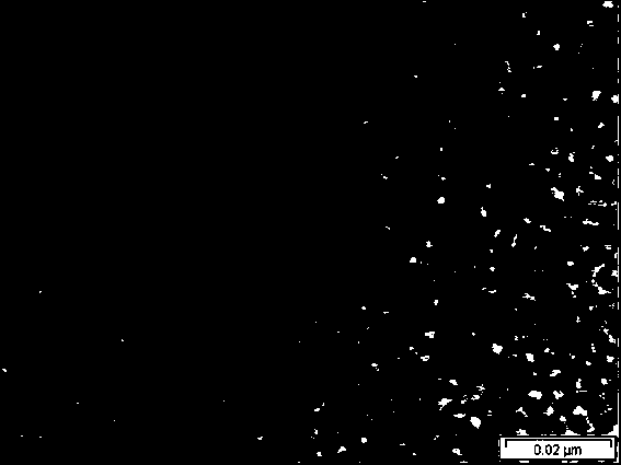
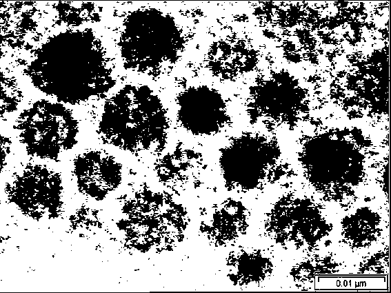
The invention relates to a strong signal and low toxicity composite nanometer material and a preparation method thereof, and belongs to the technical field of material preparation, wherein Au-Cu2S quantum dots are adopted as an inner core, and ZnS is adopted as a shell to wrap the Au-Cu2S quantum dot inner core to prepare the Au-Cu2S/ZnS quantum dots. The preparation method comprises: preparing Au nanoparticles, preparing Au-Cu2S composite nanoparticles, and preparing Au-Cu2S/ZnS quantum dots. According to the present invention, the particle size of the obtained the composite nano-particles can be controlled in several nanometers to tens of nanometers, and the obtained the composite nano-particles can be used for cytotoxicity tests and biological labeling carriers or targeted drug delivery carriers, can be provided for observing fluorescence signals through a biological imaging system, can be combined with folic acid so as to provide biocompatibility, can further enter in vivo cells and be combined with the cells so as to be adopted as the biological probe marker to perform biological labeling, and can form the durg complex, wherein the durg complex can reside on the cancer cell surface.

Description

technical field [0001] The invention belongs to the technical field of material preparation, and relates to a composite nanometer material and a preparation method thereof. Background technique [0002] Nanoparticles refer to particles with a size between 1 and 100 nm. It is a group of atoms or molecules composed of a very small number of atoms or molecules. It is a field between macroscopic substances and microscopic atoms and molecules. It is a typical mesoscopic system. Its special structure causes it to have many unique properties, such as surface effect, small size effect, quantum size effect, macroscopic quantum tunneling effect, etc. Cu 2 As a new type of P-type semiconductor nanomaterial, S has a bandgap of about 1.2eV and its unique physical and chemical properties can be used as biological probes for in vivo and in vitro detection, and has been highly valued in the fields of biology and medicine. If the surface of a single quantum dot is coated with another sem...

Claims


Application Information

Patent Timeline
IPC IPC(8): C09K11/58C09K11/02B82Y20/00B82Y30/00B82Y40/00G01N21/64A61K49/08A61K47/02A61P35/00
Inventor 刘丽炜王玥胡思怡黄晨
Owner CHANGCHUN UNIV OF SCI & TECH
Why Eureka
Try Eureka