In-situ tissue solidification engineering scaffold and preparation method thereof

A tissue engineering scaffold and in-situ curing technology, applied in the field of biomedicine, can solve the problems of low mechanical strength, unable to meet the needs of repairing load-bearing parts, etc., and achieve the effects of improved initial strength, simple preparation method and good biocompatibility

Active Publication Date: 2014-12-10
CHANGCHUN INST OF APPLIED CHEMISTRY - CHINESE ACAD OF SCI
View PDF4 Cites 2 Cited by
  • Summary
  • Abstract
  • Description
  • Claims
  • Application Information

AI Technical Summary

Problems solved by technology

However, the above technologies all use sodium salt as a porogen. After the sodium salt is injected into the tissue with t

Method used


Image

  • In-situ tissue solidification engineering scaffold and preparation method thereof
  • In-situ tissue solidification engineering scaffold and preparation method thereof
  • In-situ tissue solidification engineering scaffold and preparation method thereof

Examples


preparation example Construction

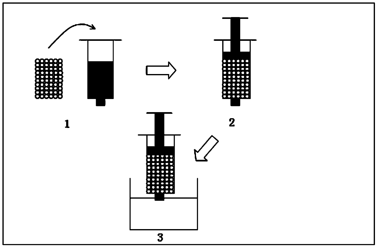
[0044] The present invention also provides a preparation method for in-situ solidified tissue engineering scaffold, comprising the following steps:

[0045] The polylactide-glycolide, biodegradable microspheres and polar organic solvents are mixed and cured in situ to obtain an in situ cured tissue engineering scaffold.

[0046] The present invention preferably mixes polylactide-glycolide (PLGA) with a polar organic solvent to obtain a PLGA solution, mixes biodegradable microspheres with the PLGA solution, and solidifies in situ to obtain in situ solidified tissue engineering stand. In order to make the obtained in-situ solidified tissue engineering scaffold applicable to bone tissue repair, the present invention preferably mixes PLGA, polar organic solvent and hydroxyapatite (HA) to obtain a PLGA solution, and mixes biodegradable microspheres with the The PLGA solution was mixed and solidified in situ to obtain an in situ solidified tissue engineering scaffold. In the prese...

Embodiment 1

[0056] Gelatin was dissolved in deionized water at 37°C to prepare a gelatin solution with a mass concentration of 25%, and then after autoclaving at 120kPa, bone morphogenetic protein (BMP-2) was added to the gelatin solution to obtain BMP-2 A gelatin solution with a concentration of 500 ng / mL.

[0057] Add 20mL of liquid paraffin and 50μL of Span-80 into the beaker, stir evenly in a water bath at 37°C, add 10mL of gelatin solution into it, stir at 200rpm for 10min, then keep the speed constant, transfer to ice at 4°C Under bath conditions, stirring was continued for 1 h to obtain a mixture.

[0058] Filter through a filter with a pore size of 300 μm, and freeze-dry the filtered microspheres at -50° C. for 24 h.

[0059] The dried gelatin microspheres are quickly and repeatedly washed with absolute alcohol to obtain the gelatin microspheres.

[0060] At 80°C, dissolve 3g of PLGA in 10g of NMP, add 1g of HA, and stir evenly to obtain a PLGA solution.

[0061] Mix 5 g of PLG...

Embodiment 2

[0067] Gelatin was dissolved in deionized water at 37°C to prepare a gelatin solution with a mass concentration of 25%, and then after autoclaving at 123kPa, bone morphogenetic protein (BMP-2) was added to the gelatin solution to obtain BMP-2 A gelatin solution with a concentration of 500 ng / mL.

[0068] Add 20mL of liquid paraffin and 50μL of Span-80 into the beaker, stir evenly in a water bath at 37°C, add 10mL of gelatin solution into it, stir at 200rpm for 10min, then keep the speed constant, transfer to ice at 4°C Under bath conditions, stirring was continued for 1 h to obtain a mixture.

[0069] Filter through a filter with a pore size of 300 μm, and freeze-dry the filtered microspheres at -40° C. for 24 h.

[0070] The dried gelatin microspheres are quickly and repeatedly washed with absolute alcohol to obtain the gelatin microspheres.

[0071] At 80°C, dissolve 3g of PLGA in 10g of NMP, add 1g of HA, and stir evenly to obtain a PLGA solution.

[0072] Mix 6g of PLGA...


PUM

PropertyMeasurementUnit
Particle sizeaaaaaaaaaa

Abstract

The invention provides an in-situ tissue solidification engineering scaffold which is formed by carrying out in-situ solidification on polylactide-glycolide and biodegradable microspheres with the existence of a polarity organic solvent. The in-situ tissue solidification engineering scaffold provided by the invention uses the biodegradable microspheres as a pore-forming agent; the microspheres in the in-situ tissue solidification engineering scaffold are gradually degraded; a pore structure of the in-situ tissue solidification engineering scaffold is gradually formed; porosity is gradually increased; and in vivo gradient pore forming from outside to inside can be implemented, so that pore forming of the in-situ tissue solidification engineering scaffold is matched with tissue penetration. In the initial stage of in-situ solidification of the in-situ tissue solidification engineering scaffold provided by the invention, due to existence of part of undegraded pore-forming agent, the initial intensity of the in-situ tissue solidification engineering scaffold provided by the invention is obviously improved. Moreover, the in-situ tissue solidification engineering scaffold provided by the invention uses the biodegradable microspheres as the pore-forming agent, has good biocompatibility and avoids damage of high permeability of particles, such as salt, to tissue cells.

Description

technical field [0001] The invention belongs to the field of biomedicine, in particular to an in-situ solidified tissue engineering scaffold and a preparation method thereof. Background technique [0002] According to statistics, there are more than 1 million patients with bone defects caused by diseases, trauma, tumors, etc. in my country every year, and the number is on the rise. Autologous bone grafting and allogeneic bone grafting are currently the main methods for treating bone defects. However, the limited source of autologous bone and the creation of new defects in the donor site limit its application. The osteoinductive ability of allogeneic bone is worse than that of autologous bone, and there is a risk of rejection and disease transmission. In recent years, more and more inorganic synthetic materials or organic synthetic polymer materials have been researched and developed as tissue engineering scaffolds or bone repair materials, and have been gradually applied c...

Claims


Application Information

Patent Timeline
IPC IPC(8): A61L27/22A61L27/20A61L27/18A61L27/50A61L27/12
Inventor 章培标张宁陈学思王宗良王宇黄晶高田林
Owner CHANGCHUN INST OF APPLIED CHEMISTRY - CHINESE ACAD OF SCI
Why Eureka
Try Eureka