Natural polymer-hydroxyapatite bone tissue engineering scaffold material with secondary three-dimensional network structure and seed-crystal induction preparation method thereof

A technology of natural polymer and hydroxyapatite, which is applied in the field of biomedical engineering scaffold materials and their preparation, can solve the problems of difficulty in realizing hydroxyapatite reconstruction, inability to discharge polymer degradation products, and difficulty in hydroxyapatite.

Inactive Publication Date: 2011-06-29
SHANDONG UNIV
View PDF2 Cites 10 Cited by
  • Summary
  • Abstract
  • Description
  • Claims
  • Application Information

AI Technical Summary

Problems solved by technology

However, since the surface of pure polymer scaffold pores is uniform and smooth, and there is no site required for crystal nucleation, hydroxyapatite is difficult to grow on it, and a hydroxyapatite layer cannot be obtained on the surface of scaffold pores

Method used


Image

  • Natural polymer-hydroxyapatite bone tissue engineering scaffold material with secondary three-dimensional network structure and seed-crystal induction preparation method thereof
  • Natural polymer-hydroxyapatite bone tissue engineering scaffold material with secondary three-dimensional network structure and seed-crystal induction preparation method thereof
  • Natural polymer-hydroxyapatite bone tissue engineering scaffold material with secondary three-dimensional network structure and seed-crystal induction preparation method thereof

Examples


Embodiment 1

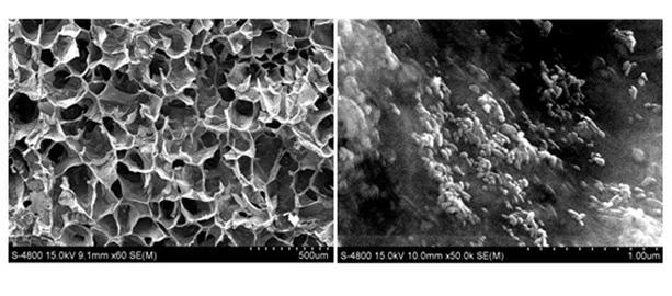
[0025] Example 1. Preparation of chitosan-hydroxyapatite secondary three-dimensional network structure bone tissue engineering scaffold material with a primary pore diameter of 50 microns and a secondary network thickness of 200 nm:

[0026] (1) Accurately weigh 1 gram of about 20 nanometers in diameter and about 120 nanometers of hydroxyapatite nanorods in length as nanoseed crystals, and disperse them in 100 milliliters of chitosan (the degree of deacetylation of chitosan is 4wt%) ≥90%, viscosity (1% solution, 20°C) ≤100 mpa.s, pH value 7.0) solution. The cross-linking agent genipin whose weight ratio of genipin to chitosan is 1:100 is added into the solution containing nanorods of hydroxyapatite, and stirred to form a sol.

[0027] (2) The sol was quickly frozen in a -80°C ultra-low temperature refrigerator, and then freeze-dried at -30°C for 36 hours to obtain a polymer porous scaffold with a primary pore diameter of 50 microns containing hydroxyapatite nanoseed crystals ...

Embodiment 2

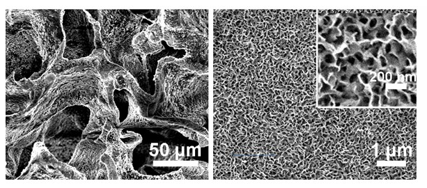
[0030] Example 2. Preparation of chitosan-hydroxyapatite secondary three-dimensional network structure bone tissue engineering scaffold material with a primary pore diameter of 100 microns and a secondary network thickness of 400 nm:

[0031] Same as Example 1, the chitosan concentration was changed to 3 wt%; the ratio of genipin to chitosan was changed to 1:200; the freeze-drying temperature was -40 o C; Mineralization time changed to 10 days;

[0032] The chitosan-hydroxyapatite secondary three-dimensional network structure bone tissue engineering scaffold material with a primary pore diameter of 100 microns and a secondary network thickness of 400 nanometers can be obtained.

Embodiment 3

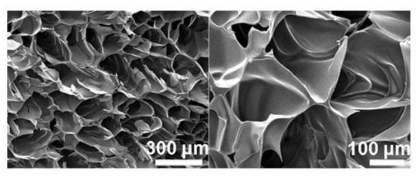
[0033] Example 3. Preparation of chitosan-hydroxyapatite secondary three-dimensional network structure bone tissue engineering scaffold material with a primary pore diameter of 150 microns and a secondary network thickness of 800 nm:

[0034] Same as in Example 1, the concentration of chitosan was changed to 2 wt%, the ratio of genipin to chitosan was changed to 1:300; the freeze-drying temperature was changed to 50° C.; the mineralization time was changed to 16 days.

[0035] The chitosan-hydroxyapatite secondary three-dimensional network structure bone tissue engineering scaffold material with a primary pore diameter of 150 microns and a secondary network thickness of 800 nanometers can be obtained.


PUM

PropertyMeasurementUnit
Wall thicknessaaaaaaaaaa
Diameteraaaaaaaaaa
Wall thicknessaaaaaaaaaa

Abstract

The invention discloses a natural polymer-hydroxyapatite bone tissue engineering scaffold material with a secondary three-dimensional network structure and a preparation method thereof. The method comprises the following steps of: mixing hydroxyapatite nanorods and a natural polymer, crosslinking by utilizing a non-toxic crosslinking agent, namely genipin, freeze-drying to prepare a porous natural polymer scaffold with nanometer seed crystals, then biomineralizing by utilizing simulation body fluid, and forming a layer of hydroxyapatite nanometer three-dimensional network structure on the walls of porous channels of the natural polymer scaffold with a micron-level three-dimensional network structure under the induction of the seed crystals. The natural polymer-hydroxyapatite bone tissue engineering scaffold material with the secondary three-dimensional network structure not only has good biocompatibility, but also is non-toxic and has high bioactivity, which indicates that the materials are expected to be widely used in repair medicine.

Description

technical field [0001] The invention relates to a biomedical engineering scaffold material suitable for repairing human hard tissue defects and its preparation, in particular to a natural polymer-hydroxyapatite secondary three-dimensional network structure bone tissue engineering scaffold material and its seed crystal induced simulated biomineral A chemical method; belongs to the technical field of biomaterials. Background technique [0002] Bone tissue engineering materials are key materials for bone repair and an important basis for the development of bone repair medicine. According to the principle of bone tissue engineering scaffold material design, this material should be a composite material prepared from degradable polymer materials and calcium-phosphorus inorganic materials, and its structure should be a multi-level network structure. Degradable polymer materials are mainly synthetic polymer materials and natural polymer materials, among which synthetic polymer mate...

Claims


Application Information

Patent Timeline
IPC IPC(8): A61L27/44A61L27/24A61L27/20A61L27/12
Inventor 刘宏王冠聪赵洪石任娜陈丽梅李建华梁小萌刘铎江怀东陶绪堂王继扬
Owner SHANDONG UNIV
Why Eureka
Try Eureka