

Tissue optical clearing agent and application thereof
A light-transparent agent and tissue technology, applied in the field of bio-optics, can solve the problems of affecting the second harmonic imaging of collagen, the destruction of collagen space structure, and the complex composition of light-transparent agent, so as to achieve uniform refractive index and endogenous The effect of strong optical signal and short time-consuming
- Summary
- Abstract
- Description
- Claims
- Application Information
AI Technical Summary
Problems solved by technology
Method used
Image

Examples
Embodiment 1
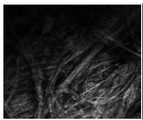
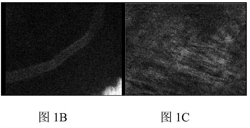
[0036] SD rats (200g) were killed by cervical dislocation after intraperitoneal injection of 1% pentobarbital 2ml, the hairs of the rats were removed with depilatory cream, the skin of the rats was cleaned with normal saline, and the skin of the back was removed with scissors. The knife removes the subcutaneous fat. After the rat skin was cleaned with phosphate buffer, soaked in 30% fructose, 10% polyethylene glycol monooctylphenyl ether (Sigma-Aldrich reagent, n=9~10) and 25% In the light clear agent aqueous solution composed of N,N,N',N'-tetra(2-hydroxypropyl)ethylenediamine, the skin becomes transparent after 15-20 minutes. Flatten the transparent skin, and use a two-photon fluorescence microscope to obtain the second harmonic signal map at a skin depth of about 120 μm (where the excitation wavelength is 900nm and the receiving wavelength is 360-540nm), to obtain Figure 1A . Use a two-photon fluorescence microscope to obtain the fluorescence signal map of skin blood vesse...
Embodiment 2
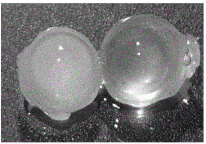
[0040] SD rats (200g) were killed by cervical dislocation after intraperitoneal injection of 1% pentobarbital 2ml, carefully took out the eyeball completely with tweezers and scissors, washed the residual blood on the eyeball with phosphate buffer, and placed it in a 4% polysaccharide POM was fixed for 24 hours, and washed with phosphate buffer after taking it out. Soak the prepared eyeballs in 70% fructose, 2% polyethylene glycol monooctylphenyl ether (Sigma-Aldrich reagent, n=9~10) and 5% N,N,N' , in the aqueous solution of light clearing agent composed of N'-tetrakis (2-hydroxypropyl) ethylenediamine, the eyeball becomes transparent after 15 to 20 minutes, such as figure 2 shown, where figure 2 The middle left picture is the eyeball before treatment, and the right picture is the transparent eyeball after treatment. Use a two-photon fluorescence microscope to obtain the second harmonic signal map of the eyeball at different depths (where the excitation wavelength is 900n...
Embodiment 3
[0042] SD rats (200g) were killed by cervical dislocation after intraperitoneal injection of 1% pentobarbital 2ml, carefully took out the eyeball completely with tweezers and scissors, washed the residual blood on the eyeball with phosphate buffer, and placed it in a 4% polysaccharide POM was fixed for 24 hours, and washed with phosphate buffer after taking it out. Soak the prepared eyeballs in 50% fructose, 5% polyethylene glycol monooctylphenyl ether (Sigma-Aldrich reagent, n=9~10) and 10% N,N,N' ,In the aqueous solution of light-transparency agent composed of N'-tetra(2-hydroxypropyl)ethylenediamine, the eyeball becomes transparent after 15 to 20 minutes, and the second harmonic signal of the eyeball at a depth of 170 μm is obtained with a two-photon fluorescence microscope Fig. (wherein excitation wavelength is 900nm, receiving wavelength is 360~540nm), obtain Figure 4 . From Figure 4 It can be seen that the second harmonic signal of the eyeball at 170 μm is clearly v...
PUM
 Abstract
Description
Claims
Application Information