
Immunofluorescence detection test strip and preparation method thereof for rapid quantitative detection of porcine epidemic diarrhea viruses

A technology for quantitative detection of porcine epidemic diarrhea, applied in antiviral immunoglobulin, botanical equipment and methods, biochemical equipment and methods, etc., achieving broad market prospects and simple operation

Active Publication Date: 2015-07-29
GUANGDONG HAID ANIMAL HUSBANDRY & VETERINARY RES INST
View PDF3 Cites 25 Cited by
  • Summary
  • Abstract
  • Description
  • Claims
  • Application Information

AI Technical Summary

Problems solved by technology

The rapid detection method of PEDV pathogen developed by anti-PEDV nanobody has not been reported at home and abroad

Method used


Image

  • Immunofluorescence detection test strip and preparation method thereof for rapid quantitative detection of porcine epidemic diarrhea viruses
  • Immunofluorescence detection test strip and preparation method thereof for rapid quantitative detection of porcine epidemic diarrhea viruses
  • Immunofluorescence detection test strip and preparation method thereof for rapid quantitative detection of porcine epidemic diarrhea viruses

Examples


Embodiment 1

[0025] Example 1 Preparation of Porcine Epidemic Diarrhea Virus Heavy Chain Single Domain Antibody

[0026] 1) Animal immunity

[0027] Multi-point inoculation of inactivated PEDV vaccine was carried out subcutaneously on the back of the neck and on both hind legs of Bactrian camels. The first immunization dose was 1 mL / head, a total of 2 heads; the second immunization was 2 mL / head after 7 days; the next immunization was 14 days apart, each immunization was 4 mL, a total of 3 immunizations, and a total of 5 immunizations in the whole process. Seven days after the last immunization, 500 mL of blood was collected to separate lymphocytes, and total RNA was extracted for the next experiment.

[0028] 2) Construction of single domain antibody library

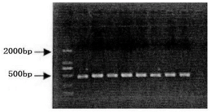
[0029] Primers were designed to amplify the VHH gene fragment, and the primers with the best effect were screened out through a large number of previous experimental studies. The sequence is as follows:

[0030] VHH-F: 5'-C...

Embodiment 2

[0046] Example 2 Preparation of PEDV polyclonal antibody

[0047] Immunize Kunming mice with PEDV inactivated vaccine, 0.5mL / mouse, a total of 5 mice, 7 days after the first immunization, the second immunization, 1mL / mouse; after that, immunize once every 14 days, 1mL / mouse, a total of 3 immunizations . Blood was collected 7 days after the last immunization to test the serum titer. If the result was positive, all the blood was separated and serum was collected as a PEDV polyclonal antibody for later use.

Embodiment 3

[0048] Example 3 Preparation of PEDV Immunofluorescence Rapid Detection Test Strip

[0049] 1) Preparation of PEDV single domain antibody

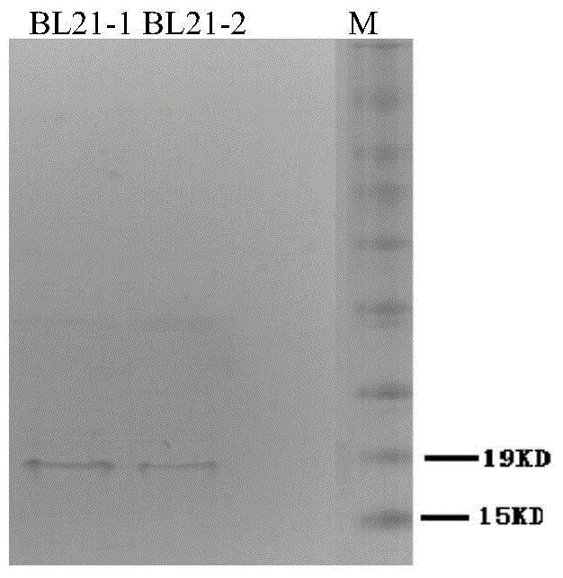
[0050] The recombinant strain BL21-1 obtained in Experimental Example 1 above was subjected to IPTG-induced expression, extraction, and purification of the target protein to obtain a PEDV single domain antibody;

[0051] 2) Preparation of PEDV single domain antibody-fluorescence microsphere probe

[0052] Carboxylation of fluorescent microspheres: Dissolve 100uL fluorescent microspheres (purchased from Guangzhou Meijintai Biotechnology Co., Ltd.) in 500 μL of purified water, then add EDC with a final concentration of 10 mg and NHS with a final concentration of 6 mg, and incubate at 27°C for 30 minutes. The reaction solution was centrifuged at 4°C at 10,000 rpm / min for 1 min, the supernatant was discarded, and repeated 3 times to remove excess EDC and NHS. The pellet was resuspended in 500 μL PBS and sonicated for 5 minutes.

[005...


PUM


Abstract

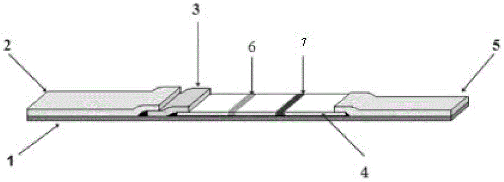
The invention discloses an immunofluorescence detection test strip and a preparation method thereof for rapid quantitative detection of porcine epidemic diarrhea viruses. The test strip comprises a sample cushion, a combination cushion, a chromatography film and a water-absorbing cushion, wherein the combination cushion is provided with a fluorescent microsphere labelled anti-PEDV (Porcine Epidemic Diarrhea Viruses) single-domain antibody; the single-domain antibody has high specificity and high sensibility against the antigen PEDV. The test strip is used for detecting PEVD viruses in breeding pig manure or PEDV pollution in feeding stuff plasma proteins on the basis of the newfound immunology principle of the specific single-domain antibody and antigen, can be used for on-site rapid detection or laboratory detection by simply taking 5-10 min; when the test strip is used in combination with a fluorescence quantitative detector, quantitative detection can be realized; the test strip is simple and convenient to operate, operators don't need professional training, a special laboratory is not needed, the limitation of the conventional detection method is overcome, and the test strip has good market prospects.

Description

technical field [0001] The invention relates to an immunofluorescence test strip for rapid quantitative detection of porcine epidemic diarrhea virus and a preparation method thereof. Background technique [0002] Porcine epidemic diarrhea (porcine epidemic diarrhea, PED) is caused by porcine epidemic diarrhea virus of Coronaviridae, an acute infectious disease invading pig intestines. Can cause vomiting, diarrhea and dehydration in pigs of all ages. Pigs of all ages are susceptible, especially for newborn piglets within 1 week of age. The morbidity rate of newborn piglets can reach 60%-100%, and the mortality rate is as high as 100%. Leading to a large number of deaths of piglets, a decline in the slaughter rate of live pigs, resulting in insufficient supply of pork in the market, affecting the stability of pork prices. The disease spreads widely in Europe and Asia, causing serious economic losses to some countries and regions every year, and bringing great harm to the pi...

Claims


Application Information

Patent Timeline
IPC IPC(8): C07K16/10C12N15/13G01N33/569G01N21/64
Inventor 贾爱卿冯晓声王贵平刘琪王悦芸周如月
Owner GUANGDONG HAID ANIMAL HUSBANDRY & VETERINARY RES INST
Learn More