A hpv-negative penile squamous cell carcinoma cell line carrying tp53 mutation and its use
A penile squamous cell carcinoma and cancer cell technology, applied in the field of HPV negative penile squamous cell carcinoma cell lines, can solve the problems of complex process, difficult to popularize and use, unclear pathogenesis, etc., and achieve the effect of stable nature
- Summary
- Abstract
- Description
- Claims
- Application Information
AI Technical Summary
Problems solved by technology
Method used
Image
Examples
Embodiment 1
[0035] Example 1. Establishment of cell lines
[0036] 1.1 Primary culture
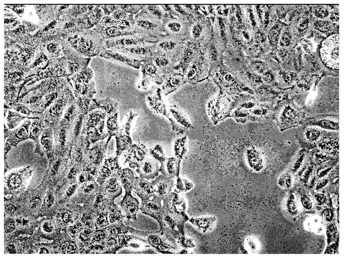
[0037] The specimens were obtained from the Sun Yat-Sen University Cancer Center, and obtained from the metastatic inguinal lymph nodes of a 41-year-old male patient with squamous cell carcinoma of the penis. After the intraoperative lymph node specimens were isolated from the body, they were separated into small tissue pieces of about 5 mm, and immediately immersed in serum-free high-glucose DMEM medium ( Add 200 U / ml penicillin, 200 μg / ml streptomycin) to the EP tube. In the biological safety cabinet dedicated to cells in the laboratory, the tumor specimens were rinsed 3 times with phosphate buffered saline (PBS), and the tumor specimens were cut into small pieces with a diameter of 1-3 mm by sterile ophthalmic scissors, and placed evenly in 25 cm. 2Add about 1-3ml high-sugar DMEM medium (adding 10% imported fetal bovine serum (FBS), 200U / ml penicillin, 200μg / ml streptomycin, used as a growth mediu...
Embodiment 2
[0042] Example 2. Immunocytochemical staining and ultrastructural morphology observation of Penl1 cell line
[0043] 2.1 Cytochemical staining
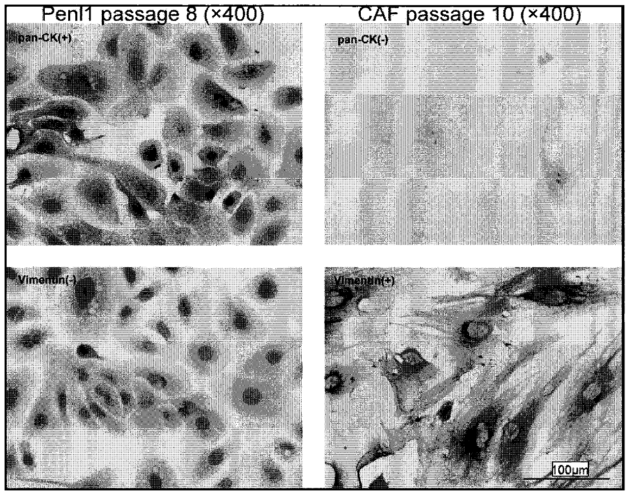
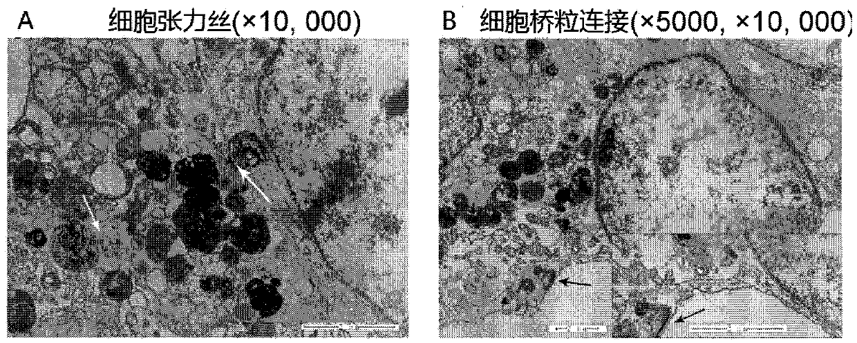
[0044] When the cells were subcultured to the 8th generation, the Penl1 cells were grown on 24mm coverslips in a six-well plate. After the cells were completely attached to the wall and in a good growth state, the six-well plate was removed from the incubator, the culture medium was removed, and PBS was used to replace the cells. Rinse the coverslip, add 4% paraformaldehyde to fix the cells for 10 minutes, use 0.3% Triton X-100 to permeabilize the cells, then block the cells with 10% goat serum for 1 hour at room temperature, and incubate pan-CK and vimentin primary antibodies at 37 degrees For 1 hour, incubate the polymerized HRP-labeled anti-mouse IgG secondary antibody at 37°C for 30 minutes, develop color with DAB for about 5 minutes, and develop color with hematoxylin for 3 minutes, dehydrate and seal the slides. The above cytoc...
Embodiment 3
[0049] Example 3. Genetic characterization of the Penl1 cell line
[0050] 3.1 Short tandem repeat (STR) cell identification
[0051] The DNA of tissue samples and cells was extracted using the AllPrepDNA / RNA / Protein Mini kit from Qigen. The DNA of metastatic lymph nodes, normal lymph nodes, and Penl1 cell line of the patient from which the samples were collected was used to amplify 19 STR loci and 1 sex-determining locus with a total of 20 loci, and the PCR amplification products were ABI3730xl electrophoresis was performed, and GeneMapper 4.0 software was used to analyze, and the STR spectrum containing 9 commonly used sites for cell line identification was compared with the three major international cell databases (American Type Culture Collection (ATCC), German Bioresource Collection (DSMZ) and Japan Cancer Resource Bank (JRCB) STR database for comparison. As shown in Table 1, the STR profile of the metastatic lymph node is consistent with that of the normal lymph node, ...
PUM
 Abstract
Description
Claims
Application Information
 - R&D
- Intellectual Property
- Life Sciences
- Materials
- Tech Scout
- Unparalleled Data Quality
- Higher Quality Content
- 60% Fewer Hallucinations
Browse by: Latest US Patents, China's latest patents, Technical Efficacy Thesaurus, Application Domain, Technology Topic, Popular Technical Reports.
© 2025 PatSnap. All rights reserved.Legal|Privacy policy|Modern Slavery Act Transparency Statement|Sitemap|About US| Contact US: help@patsnap.com


