

Method for cultivating hepatitis e virus
A technology of hepatitis E virus and virus, which is applied in the field of successfully culturing hepatitis E virus strains in vitro and improving the replication efficiency of progeny viruses, and can solve problems such as restricted replication of HEV viruses
- Summary
- Abstract
- Description
- Claims
- Application Information
AI Technical Summary
Problems solved by technology
Method used
Image

Examples
Embodiment 1
[0019] Embodiment 1: A549 cell culture
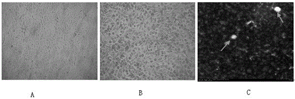
[0020] A549 cells (purchased from ATCC, USA, number CCL-185) were taken out of liquid nitrogen and quickly placed in warm water at 37°C to rapidly melt the cell suspension; then cultured in DMEM containing 10% newborn bovine serum (purchased from GIBCO Invitrogen Corporation) Adjust the pH to 7.2 and incubate at 37°C for about 4 hours. Discard all the culture medium and add fresh culture medium to continue the culture. After growing into a dense monolayer, wash the cells with PBS (purchased from GIBCO Invitrogen Corporation) and trypsin-EDTA (purchased from GIBCO Invitrogen Corporation) to digest, add the above culture medium, and place at 37°C, 5% CO 2 Continue culturing in the incubator, such as figure 1 A Normal A549 cells are shown.
Embodiment 2
[0021] Embodiment 2: Cultivate HEV with the DMEM medium that adds the pregnant woman's late serum
[0022] 1. Prepare a PBS (pH7.4) feces suspension with a weight-volume ratio of 10% from the collected HEV-positive feces for HEV isolation, shake vigorously to emulsify the feces, centrifuge at 12,000g for 10 minutes at 4°C, and collect the supernatant. 0.22μm filter membrane filter to sterilize, add double antibody solution (400U / mL penicillin and 1000U / mL streptomycin) with 2% volume of virus solution, treat at 4°C for 1h, store at -80°C for later use, and detect the virus by Real-timeqPCR The copy number is 2×10 6 Copy number / mL;
[0023] Isolated from HEVRNA-positive pig feces samples in Kunming, Yunnan, China, the genotype is type 4, and the GenBank database number is No.JF747598;
[0024] 2. Dilute the cells (A549 cells) in Example 1 by 2×10 5 Inoculate each well into a 6-well plate, and use DMEM medium containing 10% fetal bovine serum by mass percentage at 37 °C, 5% C...
Embodiment 3
[0033] Example 3: (1) HEV virus (isolated from HEVRNA-positive pig feces samples in Kunming, Yunnan, China, genotype 4, GenBank database number No. JF747598) solution was sterilized through a 0.22 μm filter membrane, and the virus was added Double antibody solution with 1% solution volume, treated at 4°C for 1 hour, and stored at -80°C for later use, the virus copy number was determined to be 2×10 by Real-timeqPCR 6 Copy number / mL, where the double antibody solution is a solution containing 400U / mL penicillin and 1000U / mL streptomycin;
[0034] (2) Divide A549 cells into 2×10 5 Inoculate each well into a 6-well plate, and use DMEM medium containing 10% fetal bovine serum by mass percentage at 37 °C, 5% CO 2 Culture the cells statically in the incubator until the cells grow into a monolayer;
[0035] (3) Inoculate 100 μL of the HEV virus suspension in step (2) into a single layer of cells, incubate at 37°C for 1 hour, and shake gently every 15 minutes to make the virus fully ...
PUM
 Abstract
Description
Claims
Application Information