A transurethral bladder ultrasound detection method, diagnostic instrument and transducer
A technology of ultrasonic transducer and ultrasonic diagnostic instrument, which is applied in the field of bladder diagnostic instrument, can solve the problems affecting the accuracy of clinical detection and low bladder resolution, and achieve the effect of improving clarity, resolution and signal-to-noise ratio
- Summary
- Abstract
- Description
- Claims
- Application Information
AI Technical Summary
Problems solved by technology
Method used
Image
Examples
Embodiment 1
[0063] Example 1: Cylindrical Array Intravesical Ultrasound Transducer
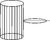
[0064] like figure 1 Shown is the schematic diagram of the intravesical ultrasound focusing transducer 11 of the present embodiment, as figure 2 As shown in its cross-sectional view, the intravesical ultrasonic focusing transducer 11 includes a plurality of ultrasonic transducing units, and each ultrasonic transducing unit includes a backing layer 111, a piezoelectric layer 112 and an acoustic matching layer 113 that are closely connected in sequence, The ultrasonic transducer units are arranged 360 degrees around the cylindrical surface, and each ultrasonic transducer is connected to a circuit, and each ultrasonic transducer unit is sequentially excited by the ultrasonic host, so as to realize 360-degree emission and reception of ultrasonic signals.
[0065] In the above embodiment, the material of the piezoelectric layer 112 can be piezoelectric ceramic material, piezoelectric thick film material, pi...
Embodiment 2
[0066] Example 2: Intravesical Ultrasound Focusing Transducer of Cylindrical Array
[0067] This embodiment adds a focusing function on the basis of Embodiment 1, which is to add a focusing unit to each ultrasonic transducing unit. The focusing transducing unit can be a mechanical structure focusing or an electronic focusing, and the mechanical structure focusing can be divided into a whole Acoustic Structure Focusing and Acoustic Lens Focusing.
[0068] like Figure 4 Shown is a schematic diagram of the focused ultrasonic transducer unit of the overall acoustic structure, wherein: the backing layer 111, the piezoelectric layer 112, and the acoustic matching layer 113 all have mechanical curved surfaces, and the radii of curvature of the three can be calculated and summed according to the requirements of the focused sound field. set up. The focus factor K is defined as the ratio of the focal length f to the transducer aperture d, ie: K=f / d. Given the focus factor K and the ...
Embodiment 3
[0071] Embodiment 3: Bladder ultrasonic diagnostic instrument
[0072] like Image 6 As shown, it is a structural schematic diagram of the bladder ultrasonic diagnostic instrument of this embodiment, which includes an ultrasonic catheter 1, a retraction / driving device 2 and an electronic imaging system 3. The front end of the ultrasonic catheter 1 is equipped with an intravesical ultrasonic transducer, and the rear end is Connect the retraction / driving device 2, the retraction / driving device 2 is connected with the electronic imaging system 3, the electronic imaging system 3 is loaded with electronic components for reconstructing images, and reconstructs the cross-sectional image and three-dimensional image of the bladder according to the received ultrasonic signal, so as to Images to judge bladder lesions. Wherein: the intravesical ultrasonic transducer is the intravesical ultrasonic transducer as described in Examples 1 and 2, where the aperture of the ultrasonic transducer...
PUM
 Abstract
Description
Claims
Application Information
 - R&D
- Intellectual Property
- Life Sciences
- Materials
- Tech Scout
- Unparalleled Data Quality
- Higher Quality Content
- 60% Fewer Hallucinations
Browse by: Latest US Patents, China's latest patents, Technical Efficacy Thesaurus, Application Domain, Technology Topic, Popular Technical Reports.
© 2025 PatSnap. All rights reserved.Legal|Privacy policy|Modern Slavery Act Transparency Statement|Sitemap|About US| Contact US: help@patsnap.com


