HPV6L1 protein-resisting antibody, preparation method therefor and application thereof

A technology of L1 protein and antibody, applied in the field of immunology, to achieve the effect of good application prospect, high affinity and high specificity

Active Publication Date: 2015-12-16
NAT VACCINE & SERUM INST
View PDF6 Cites 6 Cited by
  • Summary
  • Abstract
  • Description
  • Claims
  • Application Information

AI Technical Summary

Problems solved by technology

[0004] Since there is no more effective specific antibody against HPV6, it is necessary

Method used


Image

  • HPV6L1 protein-resisting antibody, preparation method therefor and application thereof
  • HPV6L1 protein-resisting antibody, preparation method therefor and application thereof
  • HPV6L1 protein-resisting antibody, preparation method therefor and application thereof

Examples


Embodiment 1

[0100] Preparation and screening of embodiment 1 hybridoma cells

[0101] 1. Immunogen Preparation

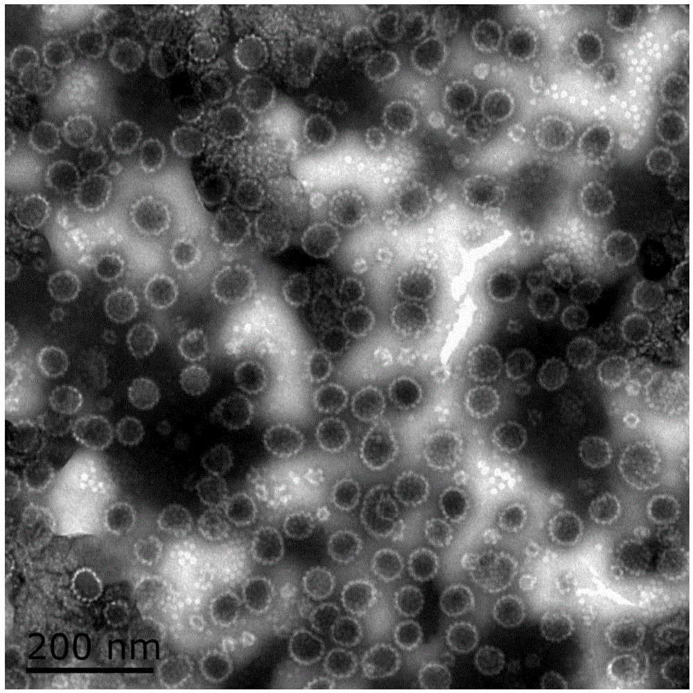
[0102] The immunogen used to prepare the monoclonal antibody of HPV6L1 protein is HPV6L1-VLP protein, and the amino acid shown in SEQ ID NO.21 is expressed through in vitro recombination according to conventional methods in the art, which can be automatically assembled into VLP, which can be seen by transmission electron microscope observation figure 1 The shown virus-like particles are spherical in diameter between 40-60 nm. The HPV6L1-VLP protein was mixed with an equal volume of complete Freund's adjuvant (CFA) or incomplete Freund's adjuvant (IFA), and fully emulsified by ultrasound to prepare the corresponding immunogen.

[0103] 2. Animal immunization

[0104] On day 0, use the immunogen mixed with CFA, inject the mice subcutaneously at multiple points on the back, and immunize 3 mice, 0.12ml / mouse, the immunogen contained is 100μg, and the injection volume of each Balb...

Embodiment 25H3

[0137] Identification of embodiment 25H3 antibody

[0138] 1. Antibody acquisition

[0139] Adult BALB / c mice were selected, and pristane was inoculated intraperitoneally, 0.5ml per mouse. After 7-10 days, the 16th passage clone5H3 hybridoma cells were inoculated intraperitoneally, 1×10 per mouse 6 -2×10 6 indivual. After an interval of 5 days, when the abdomen was obviously enlarged and the skin felt tense when touched with hands, ascites was collected with a No. 9 needle.

[0140] 2. Antibody Purification

[0141] The ascitic fluid was centrifuged at 13000 rpm / min for 30 minutes to remove cell components and other precipitates, and the supernatant was collected. Purify by ProteinG affinity chromatography and SepharoseCL-4B gel filtration, and finally obtain the monoclonal antibody clone5H3 of HPV6L1 protein, the concentration of which is above 1mg / ml.

[0142] 3. Antibody purity test

[0143] The purified antibody was subjected to 12% SDS-PAGE electrophoresis, and the...

Embodiment 34F8

[0153] The identification of embodiment 34F8 antibody

[0154] 1. Antibody acquisition

[0155] Except for inoculating clone4F8 hybridoma cells, the same method as in Example 2-1 was used.

[0156] 2. Antibody Purification

[0157] Using the same method as in Example 2-1, the monoclonal antibody 4F8 of HPV6L1 protein was finally obtained with a concentration above 1 mg / ml.

[0158] 3. Antibody purity test

[0159] Using the same method as in Example 2-1, the results showed that the purity was above 95%.

[0160] 4. Antibody class and subclass identification

[0161] Using the same method as in Example 2-1, the results show that the monoclonal antibody produced by the clone4F8 cell line belongs to IgG 2b type.

[0162] 5. Antibody light chain and heavy chain variable region gene sequence determination

[0163] The mRNA of clone4F8 hybridoma cells was extracted, reverse-transcribed into cDNA, high-fidelity PCR amplification was performed using variable region universal pr...


PUM

PropertyMeasurementUnit
Diameteraaaaaaaaaa

Abstract

The invention provides an antibody or an antigen bound part which is specifically bound to a human papilloma virus low-risk sub type-type 6 L1 protein and/or VLP. The antibody or the antigen bound part comprises CDR1, CDR2 and CDR3 regions of a heavy chain variable region shown in SEQ ID NO. 1-3 and CDR1, CDR2 and CDR3 regions of a light chain variable region shown in SEQ ID NO. 4-6. The antibody or the antigen bound part which is specifically bound with the human papilloma virus low-risk sub type-type 6 L1 protein and/or VLP has no cross reactions with most of other HPV sub types such as HPV11, 16, 18, 31, 33, 35, 39, 45, 51, 52, 56, 58, 59, 68 and the like, is high in specificity, has a characteristic of neutralizing a HPV6 pseudovirus and blocking infection of the pseudovirus, and is high in affinity. A twin-antibody sandwiched ELISA detection kit established by two antibodies can be used for detecting the presence or level of HPV6L1 protein and the antibody can be further used for treatment of patients and passive immunity of susceptible population and has a good application prospect.

Description

technical field [0001] The present invention relates to the field of immunology, and more specifically, the present invention relates to an antibody specifically binding to human papillomavirus HPV6L1 protein, a preparation method and its application in the preparation of an in vitro detection kit. Background technique [0002] Human papillomavirus (HPV) is a group of non-enveloped small DNA viruses that infect human skin and mucosal epithelial tissues, and can induce verrucous hyperplasia and even cause benign or malignant tumors. According to the difference between benign and malignant lesions induced by infection, it can be divided into high-risk HPV and low-risk HPV. High-risk HPV infection is closely related to the occurrence of cervical cancer and precancerous lesions, and is also related to the occurrence of mucosa-related cancer and precancerous lesions. Among the malignant lesions associated with high-risk HPV infection, the incidence of cervical cancer in women is...

Claims


Application Information

Patent Timeline
IPC IPC(8): C07K16/08C12N5/20G01N33/569G01N33/577
Inventor 李启明陈实张靖韩子泊郭舒扬马智静杜丽芳
Owner NAT VACCINE & SERUM INST
Why Eureka
Try Eureka