A monoclonal antibody secreting anti-wt1 protein and its application

A monoclonal antibody, antigen technology, applied in the direction of anti-animal/human immunoglobulin, anti-receptor/cell surface antigen/cell surface determinant immunoglobulin, analytical materials, etc., can solve the problem of reduced sensitivity and other problems , to achieve high specificity and sensitivity

Active Publication Date: 2018-08-17
FUZHOU MAIXIN BIOTECH CO LTD
View PDF2 Cites 0 Cited by
  • Summary
  • Abstract
  • Description
  • Claims
  • Application Information

AI Technical Summary

Problems solved by technology

In the study of the diagnostic value of various tumors, it was found that the monoclonal antibody was different from the polyclonal antibody (c19, also a DAKO product) in the discrimination of Wilms tumor, and the positive rate of 6F-H2 was 36%. %, and 47% for multiple monoclonal antibodies, the difference in the positive rate reflects the specificity of monoclonal antibody recognition epitopes, but also limits the decrease in sensitivity caused by different affinity

Method used


Image

  • A monoclonal antibody secreting anti-wt1 protein and its application
  • A monoclonal antibody secreting anti-wt1 protein and its application
  • A monoclonal antibody secreting anti-wt1 protein and its application

Examples


Embodiment 1

[0031] Example 1 Preparation of recombinant WT1 protein fragments

[0032] i. WT1 recombinant protein synthesized a 577bp WT1 gene fragment (Nanjing GenScript Biotechnology Co., Ltd. company), added at the 5' and 3' ends of the gene, respectively Eco RI and xho I restriction sites and protective bases facilitate endonuclease digestion. The pET30a-GST vector was purchased from Merck, and the vector was also used Eco RI and xho I was digested, recovered by electrophoresis, and connected with T4 DNA ligase. The ligation product was transformed into Escherichia coli competent cells BL21 codonplus (Novogen, Merck), the clones on the plate were picked and inoculated, the plasmid DNA was extracted and identified by PCR. PCR showed that the fusion protein gene positive clones were sequenced and analyzed, and the clones with completely correct sequences were adopted.

[0033] Protein Expression and Purification

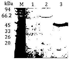
[0034] gene cloning for recombinant protein expression

[003...

Embodiment 2

[0078] Example 2 Establishment of hybridoma cell lines

[0079] 1. Immunity

[0080] About 500 μg (0.5 mg / ml) of the soluble recombinant protein obtained in Example 1 was emulsified with complete Freund's adjuvant (Sigma Company), and immunized with 4-6 week-old female Balb / c mice (purchased from Beijing Weitong Lihua Experimental Animal Technology Co., Ltd.), and each mouse was injected subcutaneously at 6 points in the abdomen, with a dose of 60 µg / only. Immunization was boosted once every 14 days, and the recombinant protein was emulsified with Freund's incomplete adjuvant (Sigma Company) at a dose of 30 μg per mouse. 7 days after the third booster immunization, indirect ELISA (wavelength 450nm) was used to detect the multi-antibody titer of the anti-immunogen in the mouse serum. 50µg / only.

[0081] 2. Cell Fusion

[0082] Aseptically prepare the mouse splenocyte suspension that reaches the immune standard, mix it with mouse myeloma cell sp2 / 0 (ATCC) at a ratio of 5:1, ...

Embodiment 3

[0087] Example 3 Preparation of monoclonal antibody by ascites induction method

[0088] 1. Ascites preparation

[0089] Cells in the logarithmic growth phase were washed with serum-free medium and suspended, counted to 5×10 5 , 1ml. The suspended cells were injected intraperitoneally into mice previously sensitized with paraffin oil. Ascites collection was started 7 days later. The removed ascites was centrifuged at 4°C at 4000 rpm for 10 min. Carefully suck out the ascites in the middle and collect in a centrifuge tube, and store at 4°C or -20°C.

[0090] 2. Purification of monoclonal antibodies

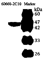
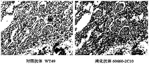
[0091] Antibody was purified from ascitic fluid by HiTrap rProtein A FF (GE Company) affinity chromatography according to the instructions. The purity was identified by SDS-PAGE gel, and the concentration was determined by Bradford method. Purified antibodies were stored at -20°C.


PUM

PropertyMeasurementUnit
melting pointaaaaaaaaaa

Abstract

The invention relates to a monoclonal antibody secreting anti-WT1 protein and its application. WT1 protein is a cell line 60660‑2C10 that can secrete IgG1 subtype monoclonal antibody through the preparation of recombinant soluble protein after analysis of WT1 protein in the present invention of Wilms, and through animal immunization and screening, its affinity constant is 3.84× 10⁹. Western blotting experiments showed that the antibody could specifically recognize recombinant WT1 protein and Wilms tumor tissue expressing WT1 protein. The antibody can be used to detect the expression level of WT1 protein in cells by immunohistochemical staining (IHC), enzyme-linked immunosorbent assay (ELISA) or western blot (Western Blot) manually or automatically, and can be used in Wilms cells Immunological diagnosis and grading of tumors, ovarian and breast cancers.

Description

technical field [0001] The invention relates to a monoclonal molecule capable of recognizing human WT1 protein molecule and a hybridoma cell line capable of secreting the antibody. Specifically, the present invention provides a monoclonal antibody against the tumor cell surface antigen WT1 molecule, which can be used for immunohistochemical staining (IHC), enzyme-linked immunosorbent assay (ELISA) or The Western Blot method is used to detect the expression level of WT1 protein in cells so as to be used in a method for diagnosing these tumors, and belongs to the field of biological detection. Background technique [0002] Wilms tumor is the most common abdominal malignant tumor, and its incidence rate ranks first among abdominal tumors in children. Tumors mainly occur within the first 5 years of life, especially between 2 and 4 years old. Wilms' tumor gene (Wilms' tumor gene, wt1 ) is a gene related to the occurrence and development of Wilms tumor. It was initially found t...

Claims


Application Information

Patent Timeline
Patent Type & Authority Patents(China)
IPC IPC(8): C07K16/30G01N33/68G01N33/577
Inventor 杨清海陈惠玲周洪辉王小亚
Owner FUZHOU MAIXIN BIOTECH CO LTD
Why Eureka
Try Eureka