Method for efficiently separating and purifying mammary epithelial cells
A technique for separation and purification of mammary epithelial cells is applied in the field of high-efficiency separation and purification of animal mammary epithelial cells in vitro to achieve the effects of less cell damage, simple and easy experimental operation, and improved culture efficiency.
- Summary
- Abstract
- Description
- Claims
- Application Information
AI Technical Summary
Problems solved by technology
Method used
Image
Examples
Embodiment 1
[0023] Example 1 Separation and Purification of Cow Mammary Epithelial Cells
[0024] 1. Sampling of breast tissue
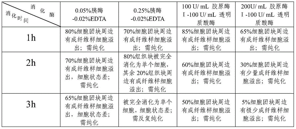
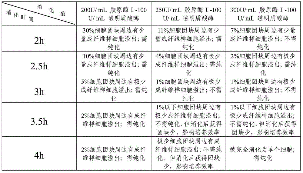
[0025] The sample was taken from an adult lactating black-and-white dairy cow in a local slaughterhouse. The mammary gland was cut open by aseptic surgery. In the parenchyma, avoid blood vessels, fat and connective tissue, and collect 8mm 3 Small and large tissue pieces were soaked in 75% alcohol for 2-3 seconds, washed in Hank’s solution containing 200 U / mL penicillin and 200 μg / mL streptomycin for about 30 seconds, incubated in the above-mentioned Hank’s solution with ice packs and brought back to the laboratory.
[0026] Trim off the visible fat and connective tissue of the tissue block in an ultra-clean workbench, wash twice with Hank’s solution containing 150 U / mL penicillin and 150 μg / mL streptomycin, and chop to 1 mm 3 Add 30mL of this Hank’s solution, shake at room temperature for 5min, discard the cleaning solution, and repeat this until the supernatan...
Embodiment 2
[0043] Example 2 Separation and purification of sheep mammary gland epithelial cells
[0044] The sample was taken from an adult late-gestation sheep, the mammary gland was aseptically cut open, and in the parenchyma, avoiding fat and connective tissue, the sample was collected 8mm 3Small and large tissue pieces were soaked in 75% alcohol for 2-3 seconds, washed in Hank’s solution containing 200 U / mL penicillin and 200 μg / mL streptomycin for about 30 seconds, incubated in the above-mentioned Hank’s solution with ice packs and brought back to the laboratory. Trim off the visible fat and connective tissue of the tissue block in an ultra-clean workbench, wash twice with Hank’s solution containing 150 U / mL penicillin and 150 Uμg / mL streptomycin, and chop to 1 mm 3 Add 30mL of this Hank’s solution to the following size, shake at room temperature for 5min, and collect about 10g of small tissue pieces for later use.
[0045] Put spare tissue pieces into 15ml of collagenase digestion...
Embodiment 3
[0046] Example 3 Separation and Purification of Yak Mammary Epithelial Cells
[0047] The sample was taken from an adult lactating yak, the mammary gland was aseptically cut open, and a 10mm sample was collected in the parenchyma, avoiding fat and connective tissue 3 Small and large tissue pieces were soaked in 75% alcohol for 2-3 seconds, washed in Hank’s solution containing 200 U / mL penicillin and 200 μg / mL streptomycin for about 30 seconds, incubated in the above-mentioned Hank’s solution with ice packs and brought back to the laboratory. Trim off the visible fat and connective tissue of the tissue block in an ultra-clean workbench, wash twice with Hank’s solution containing 300 U / mL penicillin and 300 Uμg / mL streptomycin, and chop to 1 mm 3 Add 30mL of this Hank’s solution to the following size, shake at room temperature for 5min, discard the cleaning solution, repeat this process until the supernatant is clear, and collect about 10g of small tissue pieces for later use. ...
PUM
| Property | Measurement | Unit |
|---|---|---|
| purity | aaaaa | aaaaa |
| purity | aaaaa | aaaaa |
Abstract
Description
Claims
Application Information
 - R&D
- Intellectual Property
- Life Sciences
- Materials
- Tech Scout
- Unparalleled Data Quality
- Higher Quality Content
- 60% Fewer Hallucinations
Browse by: Latest US Patents, China's latest patents, Technical Efficacy Thesaurus, Application Domain, Technology Topic, Popular Technical Reports.
© 2025 PatSnap. All rights reserved.Legal|Privacy policy|Modern Slavery Act Transparency Statement|Sitemap|About US| Contact US: help@patsnap.com


