
Preparing method of biological source venous valve

A venous valve, bio-derived technology, applied in the field of biomedical engineering, can solve the problems of not eliminating the root cause of varicose veins, unable to restore the venous valve, affecting the appearance of limbs, etc., achieving good biophysical properties, and not easy to acute immune rejection. , the effect of improving antithrombotic properties

Active Publication Date: 2016-07-20
曾祥军
View PDF10 Cites 8 Cited by
  • Summary
  • Abstract
  • Description
  • Claims
  • Application Information

AI Technical Summary

Problems solved by technology

The cause of varicose veins of the lower extremities is venous valve insufficiency, which leads to venous blood filling and high pressure in the lower extremities, resulting in varicose veins. Varicose veins cause local blood circulation disorders and affect the appearance of the limbs. Saphenous vein stripping, these methods are only symptomatic treatment, can not restore the function of the venous valve, do not eliminate the root cause of varicose veins, and cause great harm to the patient, the patient recovers slowly after surgery, the recurrence rate is relatively high, and it will cause multiple Surgical scar remnants
[0003] Biomaterials exhibit good biocompatibility and hemodynamic characteristics in the human body. There are no reports on biogenic venous valves for the treatment of varicose veins in the lower extremities.

Method used


Image

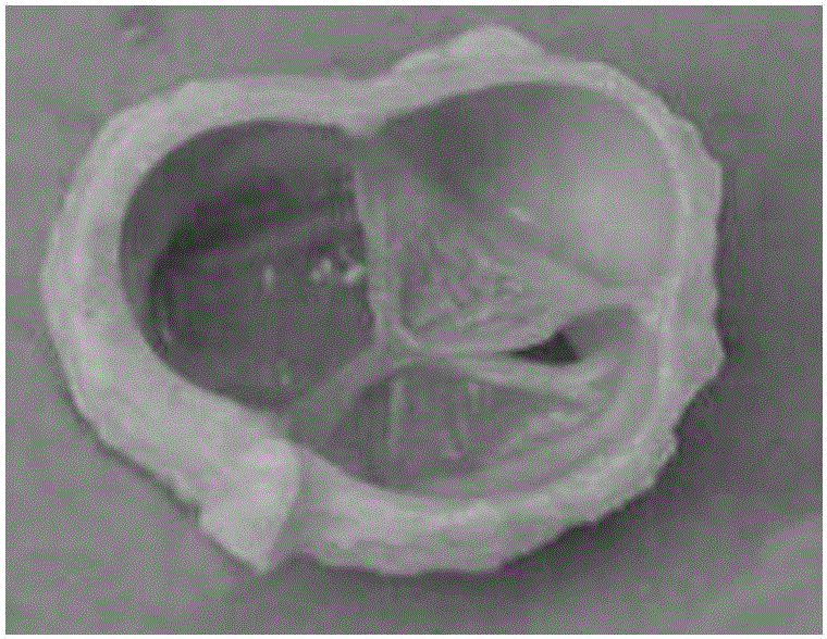
  • Preparing method of biological source venous valve
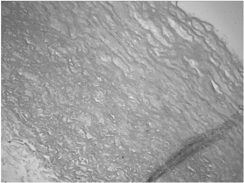
  • Preparing method of biological source venous valve
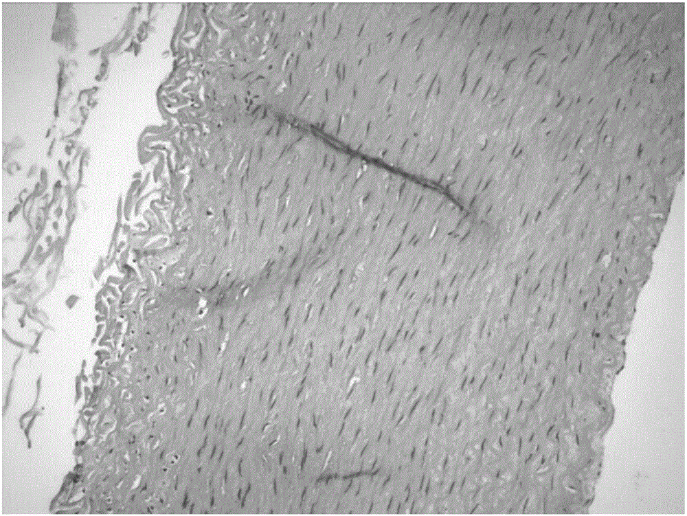
  • Preparing method of biological source venous valve

Examples


Embodiment 1

[0025] a. Take fresh bovine jugular vein pipes with a diameter of 0.5-1.5 cm and with valves slaughtered daily in slaughterhouses, turn them over and find the valve part, cut out a section with a length of 3-5 cm that contains a complete valve, turn it back and trim it Adipose tissue and excess connective tissue around the blood vessels were removed, and blood clots were removed by antegrade lavage with normal saline, then immersed in 0.1% brogeramine solution for disinfection for 30 minutes, and then rinsed with PBS solution for later use.

[0026] b. The material obtained in step a was treated with PBS solution containing 0.5% TritonX-100 for 48 hours, the cells were lysed, and washed with PBS solution. Then immersed in PBS solution containing 0.025% trypsin and 0.02% EDTA (0.025g trypsin, 0.02g EDTA dissolved in 100mL PBS solution) for 30min, digested cells, and washed with PBS solution. Finally, immerse in PBS solution containing 30u / ml DNaseI and 0.3mg / ml RNaseA for 24 ho...

Embodiment 2

[0031] The preparation process of the venous valve in this example is basically the same as that in Example 1, the difference is that the heparin binding process only includes one covalently bonded heparin treatment process described in step c, and the whole heparin binding process can be abbreviated as c. The obtained finished venous valve is stored in the same manner as in Example 1, that is, placed in 70-80% alcohol and sealed at room temperature.

Embodiment 3

[0033] The preparation process of the venous valve in this example is basically the same as in Example 1, the difference is that: 1. The time for cell lysis treatment, cell digestion and nucleic acid digestion treatment in step b is 36h, 60min and 36h respectively; 2. The heparin binding process only Including the ion-binding heparin treatment process described in step d once, the whole heparin binding process can be abbreviated as d. The obtained finished venous valve is placed in 70-80% alcohol and sealed and stored at room temperature.


PUM


Abstract

The invention belongs to the field of biomedical engineering, and particularly relates to a preparing method of a biological source venous valve.In the preparing process, a biological material is cleaned, disinfected, subjected to cell lysis, cell digestion and nucleic acid digestion and then combined with heparin, and a covalence-ion-covalence heparin combining method is adopted in the combining process.The biological source venous valve prepared through the method is high in biophysical performance, antithrombus and anti-calcification performance and has certain clinical application and popularization prospects.

Description

technical field [0001] The invention belongs to the field of biomedical engineering, and specifically refers to a preparation method of a biogenic venous valve. Background technique [0002] Varicose veins of lower extremities is a common clinical disease, which is prone to occur in people over 45 years old. Patients rarely have obvious clinical symptoms in the early stage, but in the later stage, the superficial veins of the lower limbs may be exposed, accompanied by symptoms such as acidity, sinking, swelling, pain, and fatigue. In severe cases, superficial thrombophlebitis, stasis dermatitis, Stasis ulcer, varicose vein bleeding. The cause of varicose veins of the lower extremities is venous valve insufficiency, which leads to venous blood filling and high pressure in the lower extremities, resulting in varicose veins. Varicose veins cause local blood circulation disorders and affect the appearance of the limbs. Saphenous vein stripping, these methods are only symptomat...

Claims


Application Information

Patent Timeline
Patent Type & Authority Applications(China)
IPC IPC(8): A61L27/36A61L27/50
CPCA61L27/3625A61L27/3641A61L27/3687A61L27/50A61L2430/36A61L2430/40
Inventor 曾祥军李健刘靓
Owner 曾祥军
Learn More