Porous metal cervical interbody fusion cage for directionally and slowly releasing rhBMP-2

A porous metal, rhbmp-2 technology, applied in medical science, prostheses, spinal implants, etc., can solve the problems of difficult to achieve cell attachment, growth matrix secretion and filling, unable to achieve three-dimensional hierarchical growth, etc. Effects of cell migration and mass transport, avoidance of hemorrhage, reduction of elastic modulus

Active Publication Date: 2016-07-27
PEKING UNION MEDICAL COLLEGE HOSPITAL CHINESE ACAD OF MEDICAL SCI
View PDF8 Cites 25 Cited by
  • Summary
  • Abstract
  • Description
  • Claims
  • Application Information

AI Technical Summary

Problems solved by technology

[0006] However, the 3D printed porous titanium alloy scaffold itself is not osteoinductive or osteoconductive, and the various 3D printed porous titanium alloy materials currently on the market are usually large in diameter, about 300-1500 μm in diameter, and the average diameter of cells is 10-20 μm For example, it is obviously too open, and the cells can only grow on the two-dimens

Method used


Image

  • Porous metal cervical interbody fusion cage for directionally and slowly releasing rhBMP-2
  • Porous metal cervical interbody fusion cage for directionally and slowly releasing rhBMP-2
  • Porous metal cervical interbody fusion cage for directionally and slowly releasing rhBMP-2

Examples


Embodiment 1

[0057] Example 1 Porous Titanium Alloy Cervical Intervertebral Fusion Device for Targeted and Slow Release of rhBMP-2

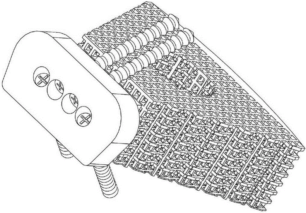
[0058] A porous titanium alloy cervical intervertebral fusion device for directional slow-release rhBMP-2. The porous titanium alloy cervical intervertebral fusion device is composed of a three-dimensional through porous structure and a solid part without a porous structure. The solid part and the three-dimensional through porous structure are integrated. The solid part seals the side of the three-dimensional penetrating porous structure away from the spinal canal; the three-dimensional penetrating porous structure is a porous titanium alloy scaffold, a three-dimensional micro-stent made of gelatin and nano-hydroxyapatite inside the porous titanium alloy scaffold, and a three-dimensional micro-stent located inside the three-dimensional micro-stent The rhBMP-2 sustained-release system consists of three-dimensional through porous structure facing the spinal cana...

Embodiment 2

[0062] Example 2 Preparation of Porous Titanium Alloy Cervical Intervertebral Fusion Device for Targeted and Slow Release of rhBMP-2

[0063] 1. Preparation of the base body of the cervical intervertebral fusion cage

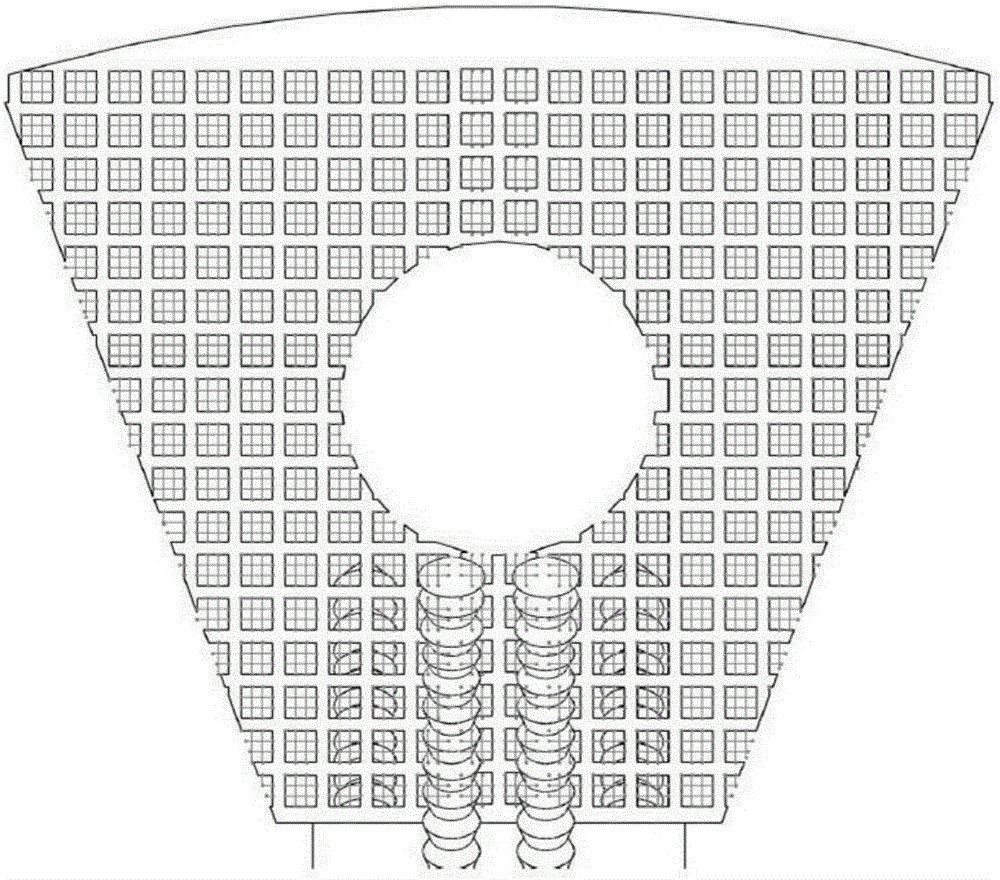
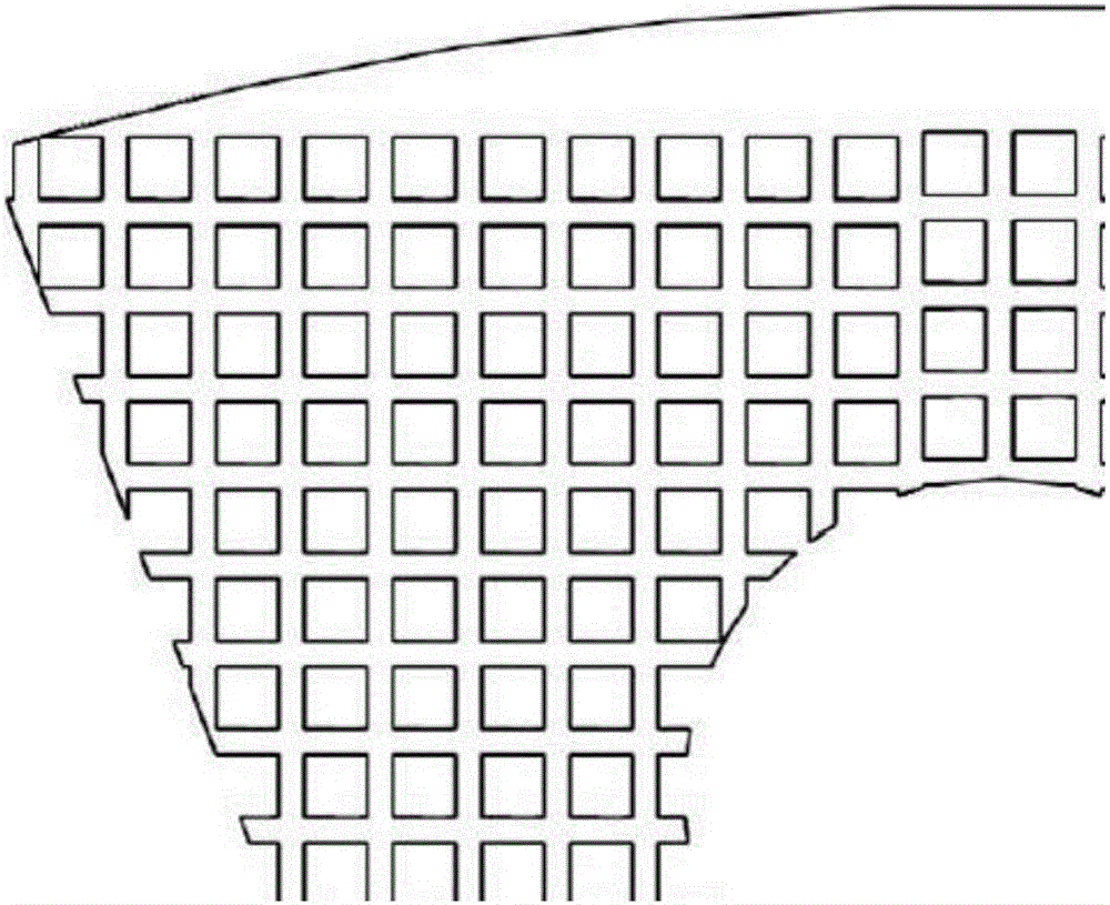
[0064] 1.1 Import the CT image into Mimics or CAD and other 3D image software to obtain a 3D image of the target bone tissue. The average pore column is 100 μm and the pore diameter is 300 μm. The image is filled and expanded with regular hexahedral structural units to obtain a personalized porous connected 3D digital image. In the model, the diameter of the middle cavity of the stent is 5 mm, the back wall is closed without holes, and the front part is a titanium plate-like structure with screw holes facing both ends.

[0065] 1.2 Using EOSM280 metal material 3D printer, titanium alloy (Ti-6Al-4V) was used as the raw material, and the basic body of the cervical intervertebral fusion cage with a solid part and a porous titanium alloy bracket was printed accordin...

Embodiment 3

[0074] Example 3 Preparation of Porous Titanium Alloy Cervical Intervertebral Fusion Device for Targeted and Slow Release of rhBMP-2

[0075] 1. Preparation of the base body of the cervical intervertebral fusion cage

[0076] 1.1 Import the CT image into Mimics or CAD and other 3D image software to obtain a 3D image of the target bone tissue. The average pore column is 100 μm and the pore diameter is 300 μm. The image is filled and expanded with regular hexahedral structural units to obtain a personalized porous connected 3D digital image. In the model, the diameter of the middle cavity of the stent is 5 mm, the back wall is closed without holes, and the front part is a titanium plate-like structure with screw holes facing both ends.

[0077] 1.2 Using EOSM280 metal material 3D printer, titanium alloy (Ti-6Al-4V) was used as the raw material, and the basic body of the cervical intervertebral fusion cage with a solid part and a porous titanium alloy bracket was printed accordin...


PUM

PropertyMeasurementUnit
Apertureaaaaaaaaaa

Abstract

The invention discloses a porous metal cervical interbody fusion cage for directionally and slowly releasing rhBMP. The cervical interbody fusion cage is manufactured through the following process that firstly, a base body of the porous metal cervical interbody fusion cage is manufactured through the 3D printing technology; then a slow release system carrying rhBMP is injected into the porous metal cervical interbody fusion cage, cooling and drying are conducted, and then the porous metal cervical interbody fusion cage can be obtained. The manufactured cervical interbody fusion cage has the elasticity modulus similar to that of a natural bone, and can directionally and slowly release rhBMP, long-term and stable bone tissue ingrowth induction is achieved accordingly, the combination problem of a bone-material interface is solved, and osseointegration capacity is improved.

Description

technical field [0001] The invention belongs to the field of spinal fusion and internal fixation, and relates to a porous metal cervical intervertebral fusion device, in particular to a porous gold cervical intervertebral fusion device capable of directional and slow release of rhBMP-2. Background technique [0002] Intervertebral disc herniation and cervical instability caused by cervical disc degeneration are common clinical spinal surgery diseases. Anterior cervical decompression and interbody fusion and internal fixation are currently the most commonly used surgical methods. However, this type of operation also faces three major problems: 1. The ACDF technique of anterior cervical plate fixation often causes postoperative swallowing discomfort for patients, and even compresses the esophagus, resulting in the possibility of esophageal leakage; 2. Bone graft does not fuse, The sinking of the titanium cage and the formation of false joints may cause fracture and displacemen...

Claims


Application Information

Patent Timeline
IPC IPC(8): A61F2/44A61L27/42A61L27/54A61L27/56
CPCA61F2/442A61F2002/4445A61L27/427A61L27/54A61L27/56A61L2300/414C08L5/02C08L5/04C08L5/08C08L5/10
Inventor 李波王海尹博苏新林黄志峰李强吴志宏邱贵兴
Owner PEKING UNION MEDICAL COLLEGE HOSPITAL CHINESE ACAD OF MEDICAL SCI
Why Eureka
Try Eureka