Biological ink material for 3D printing and preparation method and application thereof

A bio-ink and 3D printing technology, applied in additive processing, medical science, prosthesis, etc., can solve the problems of immature biological 3D soft tissue printing technology, slow cellularization and vascularization, low immunogenicity of dermal matrix, etc. , to achieve vascularization and overall performance improvement promotion, promote adhesion growth, and achieve individual targeted effects

Active Publication Date: 2016-08-24
THE FIRST AFFILIATED HOSPITAL OF SUN YAT SEN UNIV +1
View PDF1 Cites 33 Cited by
  • Summary
  • Abstract
  • Description
  • Claims
  • Application Information

AI Technical Summary

Problems solved by technology

In addition, the structure of the dermis of pigs, cattle or sheep is similar to that of humans, and the dermal matrix formed after decellularization has low immunogenicity and can be used clinically, but its internal fiber arrangement is too dense, the material has poor nutrient penetration, and the speed of cellularization and vascularization Slow, low graft survival rate, can only be used as a temporary covering for wounds, and cannot be used as a soft tissue scaffold for the construction of tissue engineering materials
[0006] To sum up, the biological 3D soft tissue printing technology in the prior art is still immature and cannot meet the clinical needs

Method used


Image

  • Biological ink material for 3D printing and preparation method and application thereof
  • Biological ink material for 3D printing and preparation method and application thereof
  • Biological ink material for 3D printing and preparation method and application thereof

Examples


Embodiment 1

[0043] The bio-ink material used for 3D printing described in this embodiment is prepared from the following raw materials by mass concentration percentage:

[0044] Biomacromolecule 5%

[0045] Water 93.5%

[0046] Coagulant 1.5%.

[0047] And the above-mentioned bio-ink materials will be washed and cross-linked to shape after printing.

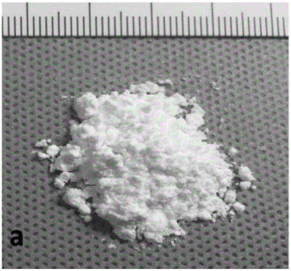
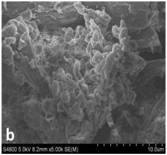
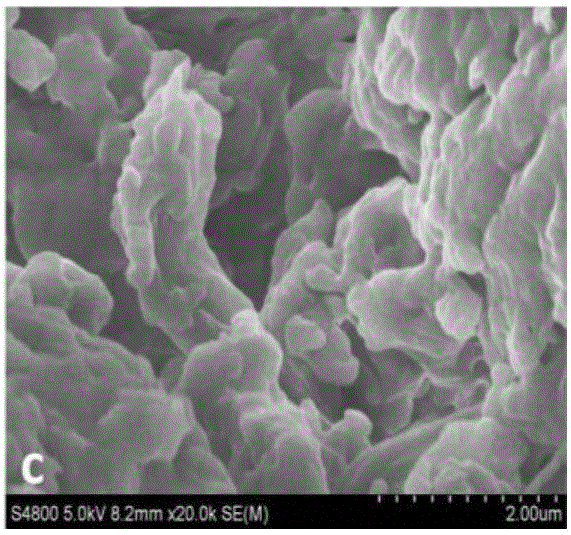
[0048] The biomacromolecule is pig dermis-derived extracellular matrix nanofiber powder.

[0049] The water is ultrapure water.

[0050]The coagulant is hydrochloric acid solution, and the molar concentration of hydrochloric acid is 3mol / L.

[0051] A preparation method for a bio-ink material for 3D printing is carried out as follows: at normal temperature, use an electronic analytical balance to weigh the biomacromolecule (i.e. pig dermis-derived ECM nanofiber micropowder) in a beaker, Add the formula amount of ultrapure water, then add a small magnetic bead, place the entire beaker on a magnetic stirrer, adjust the rotation speed to 8...

Embodiment 2

[0060] The characteristics of this embodiment are: the bio-ink material used for 3D printing described in this embodiment is prepared from the following raw materials in terms of mass concentration percentage:

[0061] Biomacromolecule 18%

[0062] Water 76%

[0063] Coagulant 6%.

[0064] And the above-mentioned bio-ink material ink will be washed and cross-linked to shape after printing.

[0065] The biomacromolecule is pig dermis-derived extracellular matrix nanofiber powder.

[0066] The water is ultrapure water. The coagulant is hydrochloric acid solution, and the molar concentration of hydrochloric acid is 3mol / L.

[0067] The crosslinking agent is glutaraldehyde solution, and the mass concentration of glutaraldehyde is 10%.

[0068] The difference in its preparation method is that the pH is adjusted to 1 during gelation, and other specific operation steps are the same as in Example 1.

Embodiment 3

[0070] The characteristics of this embodiment are: the bio-ink material for 3D printing described in this embodiment is prepared from the following raw materials in terms of mass concentration percentage:

[0071] Biomacromolecules 8.5%

[0072] water 90%

[0073] Coagulant 1.5%.

[0074] And the above-mentioned bio-ink material ink will be washed and cross-linked to shape after printing.

[0075] The biomacromolecule is pig dermis-derived extracellular matrix nanofiber powder.

[0076] The water is ultrapure water.

[0077] The coagulant is hydrochloric acid solution, and the molar concentration of hydrochloric acid is 3mol / L.

[0078] The crosslinking agent is glutaraldehyde solution, and the mass concentration of glutaraldehyde is 10%.

[0079] Other specific operation steps are the same as embodiment 1.


PUM


Abstract

The invention discloses biological ink material for 3D printing and a preparation method and application thereof. The biological ink material is prepared by, by mass concentration percentage, 5-18% of biological macromolecules, 76-93.5% of water or 76-93.5% of pre-crosslinking agent, and 1.5-6% of coagulant, wherein the sum of the mass concentration percentage of the above raw materials is 100%. The biological ink material is characterized in that water washing and crosslinking are performing for shaping after printing. The biological ink material has the advantages that the biological ink material can be easily squeezed out during printing, does not block printing needles and can be easily shaped after being squeezed out; the biological ink material is low in antigenicity in the bodies of organisms, low in caused rejection reaction, biodegradable, nontoxic to the organisms and high in safety.

Description

technical field [0001] The invention relates to a biological ink, in particular to a biological ink material for 3D printing and its preparation method and application. Background technique [0002] The cases of 3D printing technology used in clinical medicine are no longer uncommon. The combination of this technology and medical treatment mainly includes the following types: ①preoperative diagnosis and surgical rehearsal model; ②implant preparation (bones, teeth, soft tissue organs, etc.) ; ③ External medical consumables (maxillofacial shaping braces, etc.). [0003] At present, the printing technology of hard tissue scaffolds such as bones and teeth is relatively mature, but the printing of soft tissue scaffolds still needs to be further improved. [0004] Most of the existing bio-inks available for soft tissue printing are polymer hydrogels, including synthetic polymer materials and natural hydrogels such as collagen: (1) synthetic gels such as polydihydroxypropyl fumara...

Claims


Application Information

Patent Timeline
Patent Type & Authority Applications(China)
IPC IPC(8): C08L89/00C08K5/07C08K3/16C08J3/24C08J3/075A61L27/36A61L27/52A61L27/58B33Y70/00B33Y80/00B33Y10/00
CPCA61L27/3633A61L27/52A61L27/58C08J3/075C08J3/24C08K3/16C08K5/07B33Y10/00B33Y70/00B33Y80/00C08L2312/00C08L2203/02C08L2201/06C08J2389/00
Inventor 祁少海陈蕾郑永太陈永明傅炀吴序嘉
Owner THE FIRST AFFILIATED HOSPITAL OF SUN YAT SEN UNIV
Why Eureka
Try Eureka