Coronary image processing method and device

An image processing and coronary technology, applied in the field of image processing, can solve problems such as inability to diagnose and treat coronary vessels, poor treatment effect, and long treatment time

Active Publication Date: 2016-08-24
CREALIFE MEDICAL TECH BEIJING
View PDF8 Cites 6 Cited by
  • Summary
  • Abstract
  • Description
  • Claims
  • Application Information

AI Technical Summary

Problems solved by technology

[0004] However, in the process of diagnosing and treating coronary artery diseases through the coronary angiography images of patients, the obtained angiography images do not show the angiography of the coronary vessels that the iodine contrast agent does not flow through and are filled, so the doctors cannot diagnose and treat coronary artery disease

Method used


Image

  • Coronary image processing method and device
  • Coronary image processing method and device
  • Coronary image processing method and device

Examples


Embodiment 1

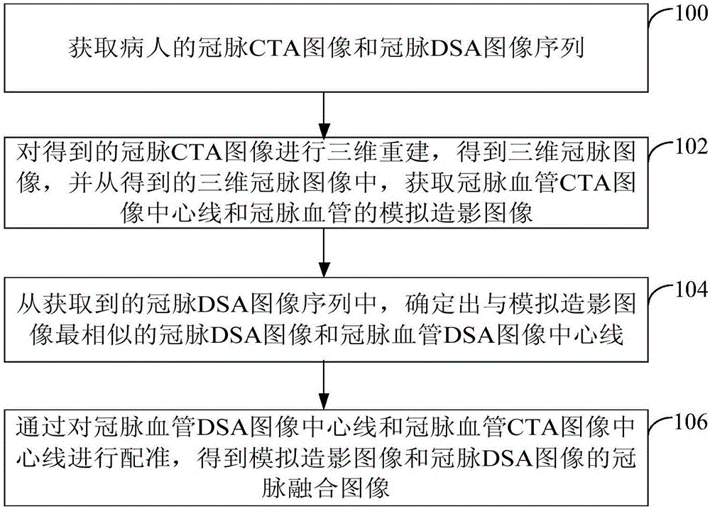
[0058] This embodiment provides a coronary image processing method. The execution subject of the embodiment of the present invention is a medical imaging system including a server, a CTA machine, and a DSA machine. The server performs data interaction with the CTA machine and the DSA machine respectively. The server sends instructions to the CTA machine and the DSA machine to obtain the patient's coronary artery CTA image and coronary artery DSA image sequence respectively, and then the server will obtain the patient's coronary artery CTA image and coronary artery DSA image sequence from the CTA machine and DSA machine respectively , and processed to obtain a coronary fusion image.

[0059] Wherein, the server can use any existing computing terminal, which will not be repeated here.

[0060] see figure 1 , the coronary image processing method proposed in the present embodiment includes the following steps:

[0061] Step 100, acquiring a coronary CTA image and a coronary DSA ...

Embodiment 2

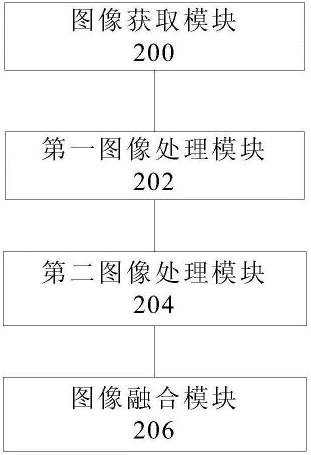
[0121] see figure 2 , the present embodiment provides a coronary image processing device for performing the above coronary image processing method, the device specifically includes:

[0122] An image acquisition module 200, configured to acquire a patient's coronary CTA image and coronary DSA image sequence;

[0123] The first image processing module 202 is configured to perform three-dimensional reconstruction on the obtained coronary CTA image to obtain a three-dimensional coronary image, and from the obtained three-dimensional coronary image, obtain a simulation of the centerline of the coronary artery CTA image and the coronary vessel Contrast images;

[0124] The second image processing module 204 is configured to determine, from the acquired coronary DSA image sequence, the coronary DSA image and the centerline of the coronary vessel DSA image most similar to the simulated angiography image;

[0125] The image fusion module 206 is configured to obtain a coronary fusio...


PUM


Abstract

The invention provides a coronary image processing method and device. The method comprises the steps of: obtaining a coronary CTA image and a coronary DSA image sequence of a patient; carrying out three-dimensional reconstruction on the obtained coronary CTA image, obtaining a three-dimensional coronary image, and from the obtained three-dimensional coronary image, obtaining a coronary blood vessel CTA image central line and a simulated contrastographic image of a coronary blood vessel; from the obtained coronary DSA image sequence, determining a coronary DSA image and a coronary blood vessel DSA image central line which are most similar to the simulated contrastographic image; and carrying out registration on the coronary blood vessel DSA image central line and the coronary blood vessel CTA image central line, and obtaining a coronary fusion image of the simulated contrastographic image and the coronary DSA image. By adopting the coronary image processing method and device, a doctor is enabled to diagnose and treat the coronary artery disease patient according to the clear coronary image.

Description

technical field [0001] The present invention relates to the technical field of image processing, in particular to a coronary image processing method and device. Background technique [0002] At present, medical imaging refers to images of human internal tissues obtained in a non-invasive manner on the human body or a certain part of the human body for medical treatment or medical research. Existing medical imaging mainly includes Computed Tomographic Angiography (Computed Tomographic Angiography, CTA) and Digital Subtraction Angiography (Digital Subtraction Angiography, DSA) and so on. When a doctor is diagnosing and treating a patient's coronary artery (coronary artery) disease, he will first obtain a coronary angiography image of the patient, and then diagnose and treat the patient's coronary artery disease through the obtained coronary angiography image. [0003] The angiography process of the existing coronary angiography image is to use DSA, first inject the iodine con...

Claims


Application Information

Patent Timeline
IPC IPC(8): G06T3/00
CPCG06T3/0068
Inventor 陈韵岱杨俊杰郭军田峰王民汉周宇李晓光
Owner CREALIFE MEDICAL TECH BEIJING
Why Eureka
Try Eureka