Method for establishing analysis module for assessing neurological functions

A technology for analyzing modules and neural functions, applied in the field of analysis modules, can solve problems such as affecting the analysis program and time, affecting the effect of the drug to be tested, and cumbersome processes.

Active Publication Date: 2016-10-05
沈孟儒
View PDF0 Cites 3 Cited by
  • Summary
  • Abstract
  • Description
  • Claims
  • Application Information

AI Technical Summary

Problems solved by technology

However, there are still many problems in the current method of using molecular markers combined with high-throughput analysis to detect neural function; for example, Huang Y et al. revealed a high content neuron-based screening in vitro to explore the A morphological computational framework that uses neuronal type III β-tubulin (TUJ1) as a molecular marker to study neuronal mechanisms (J Neurosci Methods.2010Jul 15; 190(2):299-309); Harrill JA et al Revealed a high-content image analysis to detect the effects of chemicals on the growth of primary rat cortical neuron axons and dendrites, using tubulin (β III After deducting the microtubule-associated protein 2 (MAP2) staining area (dendrites) from the tubulin) staining area (all), the remaining area represents the axon, and the growth of nerve cells is analyzed (Neurotoxicology.2013Jan; 34:61-73 ); Nieland TJ et al. used MAP2, postsynaptic density protein 95-synapsin 1 (Psd95-Syn1), and postsynaptic protein-synapsin 1 (Gphn-Syn1) to identify synaptogenesis s...

Method used


Image

  • Method for establishing analysis module for assessing neurological functions
  • Method for establishing analysis module for assessing neurological functions
  • Method for establishing analysis module for assessing neurological functions

Examples


Embodiment 1

[0031] Embodiment 1: primary cultured cells and immunofluorescent staining

[0032] Compared with the traditional method of inducing differentiation of cell lines, the method of primary culture of nerve cells is closer to the state of nerve cells, can evaluate the formation of early neural networks, and the experimental results are not affected by differentiation drugs, so the present invention preferably uses the first generation Cultured cells.

[0033] (1) Primary cultured cells

[0034] The cerebral cortex (cortex) of mice (B6mice pup) from 0 to 1 day after birth was collected, the meninges were removed, and the tissue was broken up with serum-free DMEM / F12 medium, and the volume of serum-free DMEM / F12 was supplemented to 5mL, add 0.5mL 1X trypsin (trypsin) and mix well, bathe in 37℃ water for 5 minutes, use 70μm filter membrane to infiltrate 1mL fetal bovine serum (fetal bovine serum, FBS) to filter impurities, centrifuge at 3,000rpm for 10 minutes, remove the supernatan...

Embodiment 2

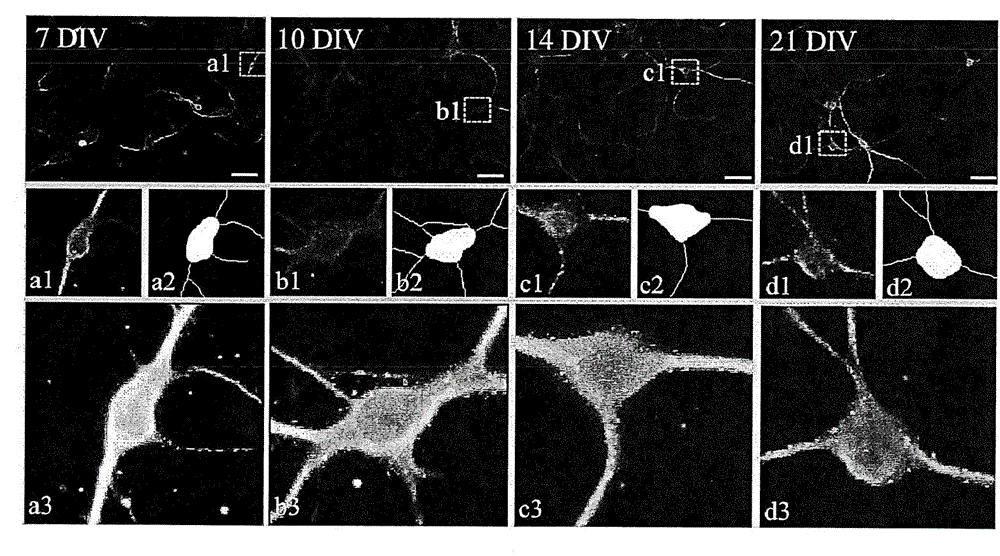
[0036] Embodiment 2: Image capture and image analysis

[0037] Fluorescence microscope imaging system was used to capture images and perform the image analysis of the above cultured cells. Three kinds of fluorescence wavelengths DAPI, FITC and Cy5 were used, and the images were automatically stored for analysis after taking pictures.

[0038] For accurate analysis of neural function, use MetaXpress 3.1 software (url ftp: / / ftp.meta.moleculardevices.com / pub / uic / software / MX31R13 / HelpDocs / MetaXpress / MetaXpress_3_1_Analysis_Guide.pdf), titled " Image Acquisition and Analysis Software (Analysis Guide), which is hereby incorporated by reference in its entirety) to design an analysis module. According to the following optimized image analysis indexes (a)-(c), a total of 32 steps for automated analysis.

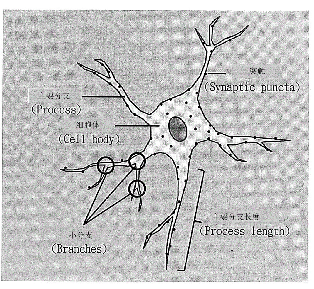
[0039] (a) Define nerve cells

[0040] Since the cultured cells contain nerve cells and glial cells, it is necessary to select the real nerve cells according to the size of the nuc...

Embodiment 3

[0083] Embodiment three: detect the impact of chemotherapeutic drugs on nerve cells

[0084] (1) Primary cultured cells

[0085] The cerebral cortex (cortex) of mice (B6mice pup) from 0 to 1 day after birth was collected, the meninges were removed, and the tissue was broken up with serum-free DMEM / F12 medium, and the volume of serum-free DMEM / F12 was supplemented to 5mL, add 0.5mL 1X trypsin (trypsin) and mix well, bathe in 37℃ water for 5 minutes, use 70μm filter membrane to infiltrate 1mL fetal bovine serum (fetal bovine serum, FBS) to filter impurities, centrifuge at 3,000rpm for 10 minutes, remove the supernatant Add the precipitated cells to the required amount of medium (medium preparation: A medium500mL, L-glutamin 100X 1.3mL, Supplement 50X 10mL / vial, Penicillin / Streptomycin 100X 2.5mL), for neuronal cell culture.

[0086] (2) Use a transparent thin-bottom microporous black plate for neuronal cell culture, and add 70 μL of poly-lysine (poly-D-lysine or poly-L-lysi...


PUM


Abstract

The invention provides a method for establishing an analysis module for assessing neurological functions. The method comprises the following steps: acquiring culture cells which include a plurality of fluorescent labels by virtue of a micro-fluorescent imaging system so as to conduct image analysis, wherein the culture cells include nerve cells and non-nerve cells; in accordance with area size of cell nucleuses and fluorescence intensity, selecting the nerve cells showing axon and dendrite fluorescent labels, eliminating non-nerve cells, and calculating the area sizes of the nerve cell bodies as well as axon and dendrite lengths and the number of branches, so that axon and dendrite growth situations of the nerve cells are determined; and in accordance with axon and dendrite fluorescent labeling ranges, calculating the number of synaptic puncta fluorescent labels on the nerve cells, so that the growth situation of the nerve cells is determined, and the neurological functions are assessed. The method disclosed by the invention can further serve as a drug screening platform, so as to rapidly detect whether drugs have a protective or neurotoxic effect on the nerve cells or not.

Description

technical field [0001] The present invention relates to a method for establishing an analysis module for evaluating nerve function, in particular to a set of rapid and representative methods for analyzing nerve growth function, including using important markers sufficient to represent nerve growth, and matching with optimized Image analysis process and program instructions; accordingly, this method can not only quickly evaluate the early nerve growth and neural network formation, but also serve as a platform for screening drugs to quickly detect the effects of drugs on nerve function. Background technique [0002] With the advancement of cancer treatment, the survival rate of patients has been gradually improved. Due to the prolongation of the patient's life expectancy, the side effects of chemotherapy drugs have been paid more attention, especially among the side effects of chemotherapy drugs, neuropathy has the greatest impact on the quality of life. Therefore, before rece...

Claims


Application Information

Patent Timeline
IPC IPC(8): C12Q1/02G01N33/569
Inventor 沈孟儒张廉筠孙苑庭张俊彦
Owner 沈孟儒
Why Eureka
Try Eureka