
In-vivo platelet instant label-free detection system and detection method

A detection system and detection method technology, applied in the directions of diagnostic recording/measurement, blood characterization device, medical science, etc., can solve the problem that real-time, full-time and instant detection cannot be realized, and achieve the effect of avoiding waiting time and convenient detection.

Pending Publication Date: 2016-10-26
MAGASSIST INC
View PDF8 Cites 1 Cited by
  • Summary
  • Abstract
  • Description
  • Claims
  • Application Information

AI Technical Summary

Problems solved by technology

[0006] However, the above-mentioned tests are all in vitro tests or rely on biomarkers for detection, which cannot achieve real-time, full-time and instant detection

Method used


Image

  • In-vivo platelet instant label-free detection system and detection method
  • In-vivo platelet instant label-free detection system and detection method
  • In-vivo platelet instant label-free detection system and detection method

Examples


Embodiment 1

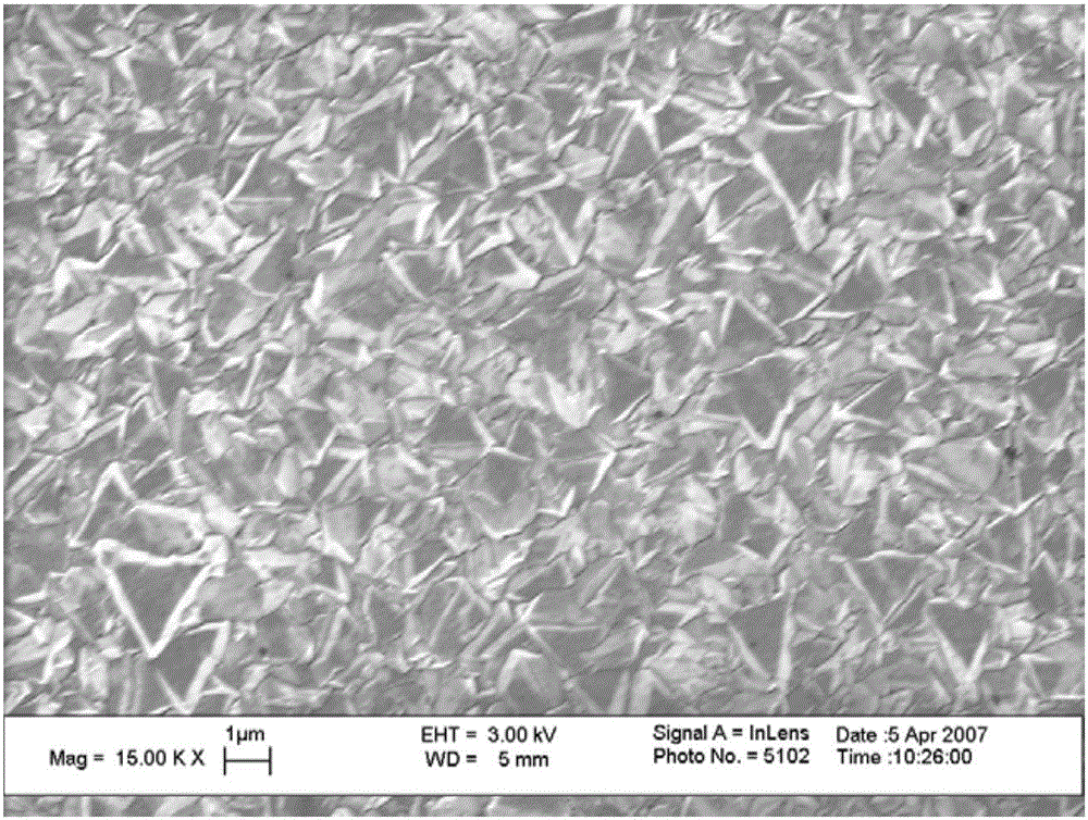
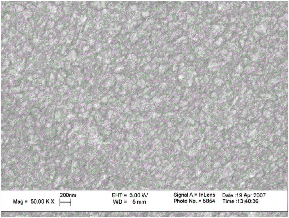
[0063] Taking the surface of an artificial heart assist device made of a titanium-based alloy as an example, a diamond film deposition process is used on the surface to form a diamond MEMS sensor on the surface of a titanium-based alloy, and to optimize its geometric distribution design on the surface of the artificial heart.

[0064] Then, the in vitro simulation experiment was carried out, and the artificial heart was connected with the dynamic blood flow simulator to simulate the flow field after the artificial heart intervention. The experiment uses donor blood for testing, and the output results are compared in two stages, the first is to compare with the computational fluid dynamics simulation results with the thrombus prediction module, and the second is to compare the monitoring results at different time points with the fluorescence microscope observation The microthrombosis distribution on the surface was compared. This checks the sensitivity of the sensor surface and...

Embodiment 2

[0067] In this embodiment, the diamond MEMS sensor is prepared on the surface of the vascular stent or the artificial blood vessel, and is used to detect the surface blood flow velocity.

[0068] The MEMS electrodes form an electric field on the diamond surface, and the surface electric field will also change under different blood flow conditions, which will be fed back to the diamond MEMS sensor to obtain a change in the electrical signal. Such as Figure 6 Shown is the current density kinetic curve of the thrombus formation process in vivo (dots are data points). It can be seen that as time goes by, platelets gradually aggregate to form blood coagulation, and the current density gradually increases.

[0069] By analyzing the changes of electrical signals and the regional concentration information of red blood cells and plasma through a fixed algorithm, the blood flow velocity on the surface of the device can be obtained in real time, so as to obtain the space of the coagulat...


PUM


Abstract

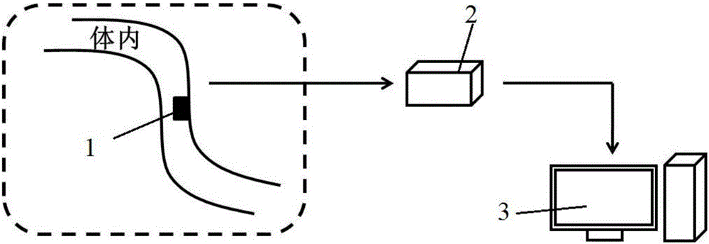
The invention discloses an in-vivo platelet instant label-free detection system. The detection system comprises a signal acquisition unit, a signal receiving unit and a signal processing unit, wherein the signal acquisition unit comprises a diamond MEMS sensor which is arranged in vivo; the diamond MEMS (micro-electro-mechanical system) sensor comprises a PC diamond thin film and electrodes which are arranged on the PC diamond thin film and are formed by conductive diamonds; the signal acquisition unit is used for acquiring kinetics signals of electrode process of platelet near-wall time-space distribution; the signal receiving unit is connected to the signal acquisition unit and is arranged in vitro, and the signal receiving unit is used for receiving the signals acquired by the signal acquisition unit; and the signal processing unit is connected to the signal receiving unit and is arranged in vitro, and the signal processing unit is used for quantitative characterization of platelet time-space distribution in accordance with the signals received by the signal receiving unit. The detection system and the detection method disclosed by the invention, which adopt in-vivo detection, can avoid an operation of extracting blood for in-vitro detection, so that the detection is facilitated; through label-free detection, the detection system and the detection method are safe and nondestructive to the blood; and through instant detection, the problem of an existing detection method which is asynchronous in waiting time as well as detection and result acquisition.

Description

technical field [0001] The invention relates to the technical field of medical detection equipment, in particular to a real-time label-free detection system and detection method for platelets in vivo. Background technique [0002] With the increasing demand for medical implants, more and more attention has been paid to the coagulation problems and acute thrombus caused by implants, and the early prediction, diagnosis and treatment of thrombus are of great significance and necessity. From the perspective of implant thrombosis mechanism, the main coagulation risk comes from the interaction between blood and implant surface, blood and flow field. The causes of thrombus formation on the surface of implants are mostly caused by local hydrodynamic factors. The main factors include the surface structure and geometry of implant materials, blood and flow field, and the feedback effect on the flow field after thrombus formation. On the other hand, due to the formation of non-physiolo...

Claims


Application Information

Patent Timeline
IPC IPC(8): A61B5/145A61B5/00
CPCA61B5/14503A61B5/6847A61B5/686A61B5/6862A61B5/6869A61B5/6876
Inventor 杨磊徐博翎陈涛赖跃坤王鲁宁刘慧玲
Owner MAGASSIST INC
Learn More