Gold-as-core silver-as-shell ''Raman silent zone'' substrate and preparation method and application thereof

A Raman silent zone and Raman substrate technology, applied in Raman scattering, material excitation analysis, etc., to achieve the effect of great clinical application prospects

Active Publication Date: 2016-12-21
FUDAN UNIV
View PDF6 Cites 15 Cited by
  • Summary
  • Abstract
  • Description
  • Claims
  • Application Information

AI Technical Summary

Problems solved by technology

However, the application of surface-enhanced Raman scattering imaging in cell imaging, especially the "Raman silent zone" of cells is still relatively small.
Although it has a bright

Method used


Image

  • Gold-as-core silver-as-shell ''Raman silent zone'' substrate and preparation method and application thereof
  • Gold-as-core silver-as-shell ''Raman silent zone'' substrate and preparation method and application thereof
  • Gold-as-core silver-as-shell ''Raman silent zone'' substrate and preparation method and application thereof

Examples


Embodiment 1

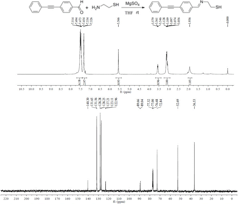
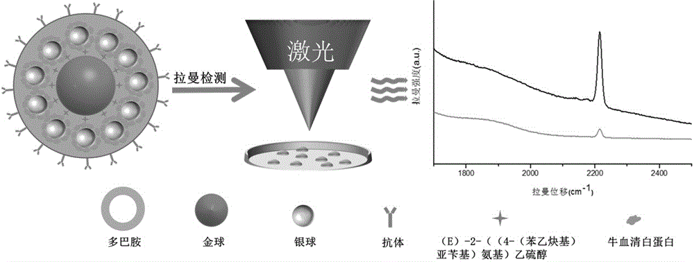
[0024] Example 1: Synthesis of a surface-enhanced Raman scattering cell imaging substrate with gold as the core and silver as the shell modified by human epidermal growth factor receptor antibody

[0025] Pipette 1.212 mL of 10 mg / mL perchlorauric acid solution and 98.788 mL of deionized water into a 250 mL three-necked flask, and add 10 uL of 10 mM (E)-2-((4-(phenylethynyl )benzylidene)amino)ethanethiol, mix it evenly, heat it in an oil bath at 140°C, and control it to reflux about once per second, then add 750 uL of 1% citric acid tris Sodium, maintain reflux for about 30 minutes, cool to room temperature, store it in a clean glass bottle, and store it at 4°C. Take the synthesized gold nanoparticles with (E)-2-((4-(phenylethynyl)benzylidene)amino)ethanethiol molecule, centrifuge it at 8000rpm for 5min, absorb the supernatant, Redissolve in borax solution, add 10 uL of 10 mM (E)-2-((4-(phenylethynyl)benzylidene)amino)ethanethiol to it, mix well and add 5 uL of 0.1 M anti Re...

Embodiment 2

[0026] Example 2: Application of Raman material with gold as core and silver as shell to identify glioma cell U251 and perform Raman imaging on it

[0027] First add about 10 5 After culturing for 12 hours, take 10-30 μL of antibody and gold as the nucleus and silver as the surface-enhanced Raman scattering substrate of the silent zone as the shell, add it to a glass dish, and then put the glass culture dish at room temperature, and Cells were incubated for 1-2 h, then aspirated the medium, washed three times with PBS, reacted with 4% paraformaldehyde solution in a 37°C incubator for 15 minutes, and finally washed three times with PBS, and added 100 uL of PBS to the Store in a refrigerator at 4°C for surface-enhanced Raman scattering imaging.


PUM


Abstract

The invention belongs to the technical field of separation and analysis of biological macromolecules, and particularly relates to a gold-as-core silver-as-shell ''Raman silent zone'' surface enhancement Raman scattering substrate and a preparation method and application thereof in Raman imaging in living cells. The preparation process is as follows: gold nanoparticles containing ''Raman silent zone'' probe molecules are in situ synthesized by hydrothermal method, the gold nanoparticles are re-dissolved in a borax solution, and ''Raman silent zone'' molecules (E) 2 ((4 (phenyl ethynyl) benzylidene) amino) ethyl mercaptan are added; ascorbic acid and silver nitrate are added for in situ reduction of silver nanoparticles; bovine serum albumin is added, and finally through dopamine self polymerization, an antibody is connected to the material for preparing the ''Raman silent zone'' surface enhancement Raman scattering substrate capable of specifically recognizing tumor cells. Experiments show that the ''Raman silent zone'' surface enhancement Raman scattering substrate has a prominent enhancing effect on a probe signal, the ''Raman silent zone'' surface enhancement Raman scattering substrate material is free of template, low-cost and low-toxicity, is used in imaging in living cells, and is novel, convenient, practical and efficient.

Description

technical field [0001] The invention belongs to the technical field of separation and analysis of biological macromolecules, and specifically relates to a surface-enhanced Raman scattering substrate of a cell "Raman silent zone" with gold as the core and silver as the shell, its preparation method and its application in cell Raman imaging. technical background [0002] Surface-enhanced Raman scattering is a special Raman spectral phenomenon, which is manifested on the rough surface of some gold, silver, and other metals. The Raman scattering signal of the molecule to be measured is affected by the surface plasmon resonance (LSPR) Geometric multiple (10 3 -10 9 )enlarge. This phenomenon greatly increases the sensitivity of Raman spectroscopy. At the same time, its smaller slit width and better spectral shape are currently widely used in high-sensitivity analysis research such as food safety, environmental monitoring, biology, medicine, and archaeology. In addition, because...

Claims


Application Information

Patent Timeline
IPC IPC(8): G01N21/65
Inventor 高明霞陈萌张玲张祥民
Owner FUDAN UNIV
Why Eureka
Try Eureka