Cell line sourced from later-stage gastric signet ring cell cancer, and establishing method and application thereof
A cell line, cancer cell technology, applied in the direction of tumor/cancer cell, microorganism-based method, animal cell, etc. The effect of stable traits, good uniformity and strong clonogenic ability
- Summary
- Abstract
- Description
- Claims
- Application Information
AI Technical Summary
Problems solved by technology
Method used
Image
Examples
Embodiment 1
[0044] Example 1 Establishment of gc-006-03 of gastric signet ring cell cancer cell line
[0045] The ascites of patients with gastric signet ring cell carcinoma was extracted, and the cells were separated by 1.077g percoll solution. After 1640 culture medium (containing 10% fetal bovine serum, 10mM HEPES, 100U / ml penicillin G, 100μg / ml streptomycin sulfate and 0.25μg / ml amphotericin B), place at 37°C, 5% CO 2 The gastric signet ring cell carcinoma cell line gc-006 was established by in vitro cell subculture in an incubator.
[0046] To establish cell lines: we firstly serially dilute the cells of the gc-006 cell line, inoculate 100-2000 cells / dish in five 10cm culture dishes, use 1640 medium (same as above), place at 37°C, 5% CO 2 Cultured in the incubator for 7-10 days. At this time, the cells are scattered and energetic monoclonal. Then select the monoclonal cells accurately, no need to add trypsin to pick the clones, and directly select the monoclonal cells with the ...
Embodiment 2
[0047] Reality Example 2 Detection of biological characteristics of gastric signet ring cell carcinoma cell line gc-006-03
[0048] 1. Cell morphology:
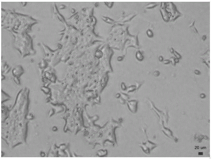
[0049] (1) Observation of live cells: After the cells have been subcultured and grown stably, they are observed under a phase-contrast microscope. The cells were adhered to the wall and overlapped, without contact inhibition ( figure 1 ).
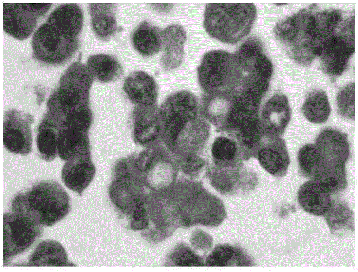
[0050] (2) HE staining: the cells were passed to 31 passages, and the number of cells reached 10 6 -10 7 Then, paraffin-embedded sections were carried out and observed under the HE staining microscope. The cells showed a malignant epithelioid shape, with a large ratio of nucleoplasm to cytoplasm, multiple nucleoli, and cells with nuclei of different sizes. Mitosis was easily seen, and a certain number of signet ring cells could still be seen ( figure 2 ).
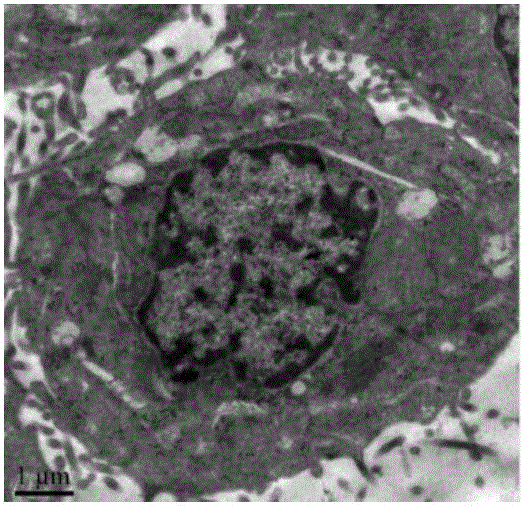
[0051] (3) Observation by transmission electron microscope: few ribosomes in cells, rich in rough endoplasmic reticulum, cyst...
PUM
 Abstract
Description
Claims
Application Information
 - R&D
- Intellectual Property
- Life Sciences
- Materials
- Tech Scout
- Unparalleled Data Quality
- Higher Quality Content
- 60% Fewer Hallucinations
Browse by: Latest US Patents, China's latest patents, Technical Efficacy Thesaurus, Application Domain, Technology Topic, Popular Technical Reports.
© 2025 PatSnap. All rights reserved.Legal|Privacy policy|Modern Slavery Act Transparency Statement|Sitemap|About US| Contact US: help@patsnap.com


