

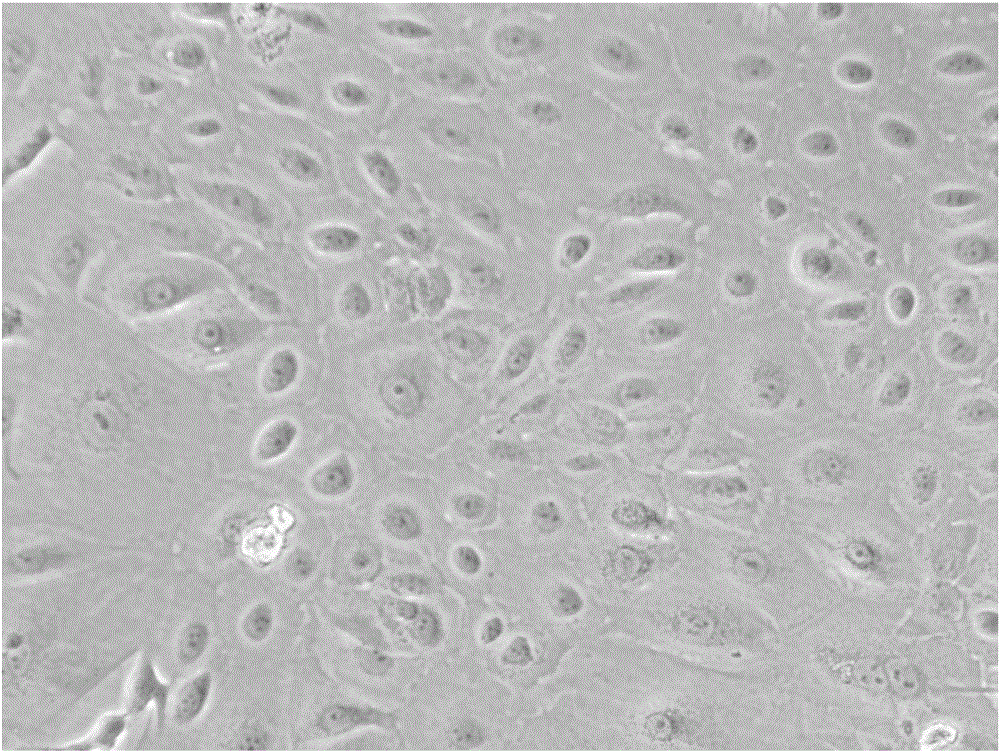
Method for preparing human amniotic membrane epithelial cells from human placenta amnion and application thereof
A technology of epithelial cells and human amniotic membrane, applied in the field of stem cell therapy, can solve the problems of high pollution rate of human amniotic membrane collection, poor cell activity, affecting cell separation and culture, etc.
- Summary
- Abstract
- Description
- Claims
- Application Information
AI Technical Summary
Problems solved by technology
Method used
Image

Examples
Embodiment 1
[0076] Example 1: Preparation of human amniotic epithelial cells from placental amniotic membrane
[0077] (1) Take out the placenta from the placenta sample collection box, place it in a white porcelain dish, use a basic balanced salt solution (KH2PO4 of 0.4g, KH2PO4 of 0.06g, Na2HPO4.12H2O of 0.132g, NaCl of 8g, 0.35g of NaHCO3, the D-glucose of 1.0g, the streptomycin of 0.10g, the penicillin of 0.06g are dissolved in water and be settled into the solution of 1L, obtain) repeatedly wash the surface, and the placenta is disinfected;
[0078] (2) Slowly tear the outer layer of amniotic membrane with surgical forceps and place it in a 150mm glass dish, use basic balanced salt solution to repeatedly clean the surface of the blood, and cut it into small pieces of amniotic membrane tissue with a diameter of 5mm;
[0079] (3) Small pieces of amniotic membrane are evenly pasted on a 100mm petri dish with the amniotic membrane epithelium facing down. After drying for 30-60 minutes ...
Embodiment 2
[0083] Example 2: Preparation of human amniotic epithelial cells from placental amniotic membrane
[0084] (1) Take out the placenta from the placenta sample collection box, place it in a white porcelain dish, use a basic balanced salt solution (KH2PO4 of 0.4g, KH2PO4 of 0.06g, Na2HPO4.12H2O of 0.132g, NaCl of 8g, 0.35g of NaHCO3, the D-glucose of 1.0g, the streptomycin of 0.10g, the penicillin of 0.06g are dissolved in water and be settled into the solution of 1L, obtain) repeatedly wash the surface, and the placenta is disinfected;
[0085] (2) Slowly tear the outer layer of amniotic membrane with surgical forceps and place it in a 150mm glass dish, use basic balanced salt solution to repeatedly clean the surface of the blood, and cut it into small pieces of amniotic membrane tissue with a diameter of 5mm;
[0086] (3) Small pieces of amniotic membrane are evenly pasted on a 100mm culture dish with the amniotic membrane epithelial layer facing down. After drying for 30-60 ...
Embodiment 3
[0090] Example 3: Preparation of human amniotic epithelial cells from placental amniotic membrane
[0091] (1) Take out the placenta from the placenta sample collection box, place it in a white porcelain dish, use a basic balanced salt solution (KH2PO4 of 0.4g, KH2PO4 of 0.06g, Na2HPO4.12H2O of 0.132g, NaCl of 8g, 0.35g of NaHCO3, the D-glucose of 1.0g, the streptomycin of 0.10g, the penicillin of 0.06g are dissolved in water and be settled into the solution of 1L, obtain) repeatedly wash the surface, and the placenta is disinfected;
[0092] (2) Slowly tear the outer layer of amniotic membrane with surgical forceps and place it in a 150mm glass dish, use basic balanced salt solution to repeatedly clean the surface of the blood, and cut it into small pieces of amniotic membrane tissue with a diameter of 5mm;
[0093] (3) Small pieces of amniotic membrane are evenly pasted on a 100mm petri dish, with the amniotic membrane epithelial layer facing down, and after drying for 30-...
PUM
 Abstract
Description
Claims
Application Information