Fibrous-protein-modified open-tubular column and application to monoclonal antibody isomer separation

A monoclonal antibody and fibrin technology, which is applied in the field of instrument analysis, can solve the problem of non-uniformity of monoclonal antibodies, and achieve the effects of improving separation effect, inhibiting adsorption, and mild preparation conditions.

Active Publication Date: 2017-03-29
SOUTH CHINA NORMAL UNIVERSITY
View PDF1 Cites 0 Cited by
  • Summary
  • Abstract
  • Description
  • Claims
  • Application Information

AI Technical Summary

Problems solved by technology

Although these changes are small, they can cause monoclonal antibodies to exhibit various types of heterogeneity, resulting in different types of variants, which differ in physicochemical properties such as molecular weight, hydrophobicity, charge, etc.

Method used


Image

  • Fibrous-protein-modified open-tubular column and application to monoclonal antibody isomer separation
  • Fibrous-protein-modified open-tubular column and application to monoclonal antibody isomer separation
  • Fibrous-protein-modified open-tubular column and application to monoclonal antibody isomer separation

Examples


Embodiment 1

[0031] A fibrin-modified open-tubular column prepared by the following steps:

[0032] (1) Rinse the open column with sodium hydroxide solution (1M), deionized water, hydrochloric acid solution (1M) and deionized water in sequence, and dry it;

[0033] (2) Mix fibrinogen solution (2 mg / mL, 500 μL), thrombin solution (0.12 μM, 25 μL) and phosphate buffer (10 mM, pH 7.4, containing 475 μL of 0.15M sodium chloride), and quickly Inject it into a pretreated open-tubular column, store it in a thermostat at 37°C for 5 hours, then flush out excess reaction solution with nitrogen and dry it in an environment at 40°C under its protection. Finally, the column was washed with 10 mM phosphate (pH value 7.4) and deionized water, and dried under nitrogen protection at 40°C to obtain a fibrin-modified open-tube column.

Embodiment 2

[0035] The application of the fibrin modified open-tubular column prepared in Example 1 in the separation of 6 cetuximab isomers comprises the following steps:

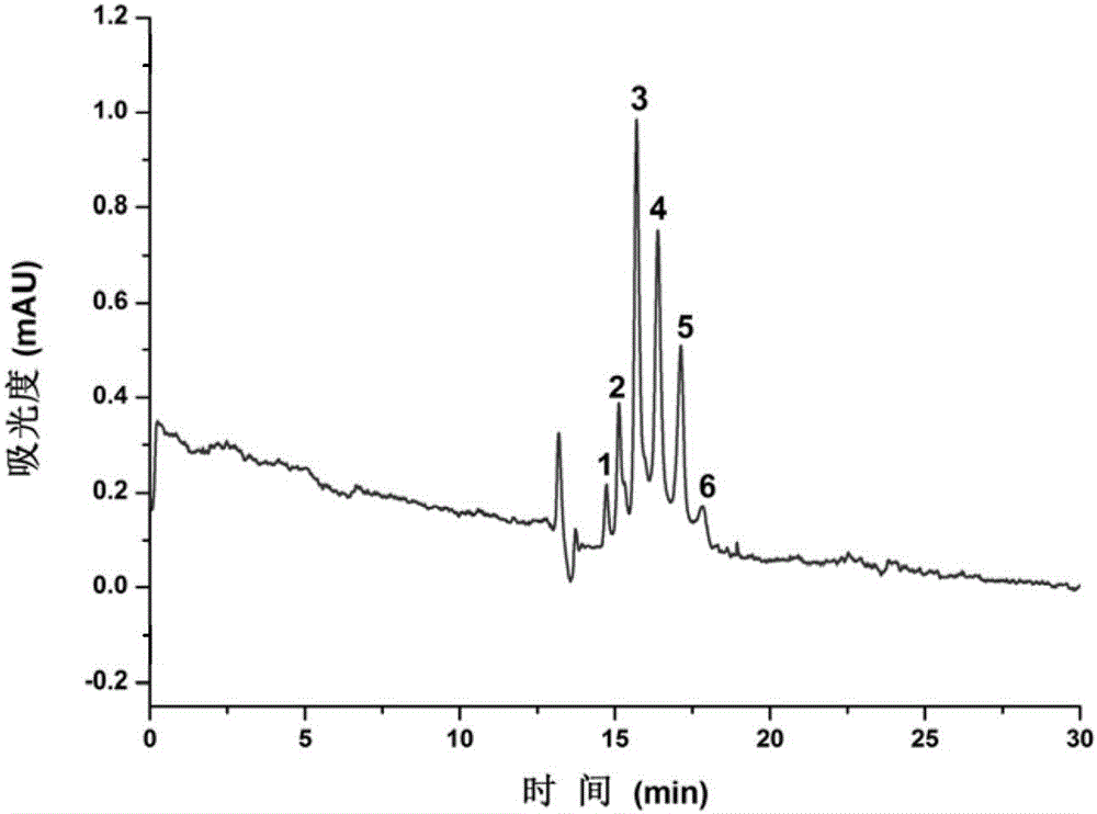
[0036] Dissolve cetuximab after centrifugation and dilution in water, and separate it by electrochromatography. The results are shown in figure 1 . The separation conditions are as follows: the inner diameter of the chromatographic column is 50 μm, the total length is 62.5 cm, the effective length is 37.0 cm, the mobile phase is 50 mM (pH 7.0) phosphate buffer, the separation voltage is 20 kV, the detection wavelength is 214 nm, and the sampling method is height Injection.

[0037] Depend on figure 1 It can be seen that the six different forms of isomers of cetuximab are well separated on the fibrin-modified open-tubular column, and the peak shape is sharp, indicating that the coated column can be effectively applied to cetuximab Identification and separation of mAb isoforms.

Embodiment 3

[0039] The application of the fibrin modified open-tubular column prepared in Example 1 in separating 5 kinds of rituximab isomers comprises the following steps:

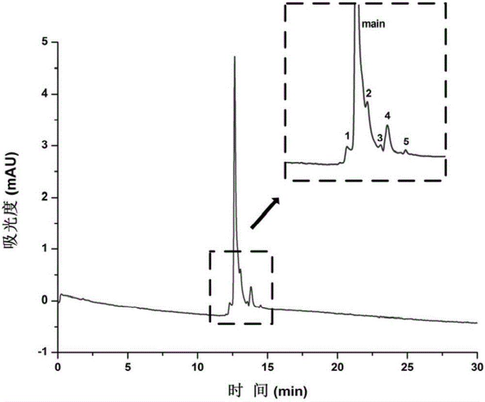
[0040] The centrifuged and diluted rituximab was dissolved in water and separated by electrochromatography. The results are shown in figure 2 . The separation conditions are as follows: the inner diameter of the chromatographic column is 50 μm, the total length is 62.5 cm, the effective length is 37.0 cm, the mobile phase is 50 mM (pH 7.0) phosphate buffer, the separation voltage is 20 kV, the detection wavelength is 214 nm, and the sampling method is height Injection.

[0041] Depend on figure 2 It can be seen that five different isomers of rituximab were successfully separated from the main peak, indicating that the coated column can be effectively applied to the identification and separation of rituximab isomers.


PUM

PropertyMeasurementUnit
concentrationaaaaaaaaaa

Abstract

The invention discloses a fibrous-protein-modified open-tubular column and an application of the fibrous-protein-modified open-tubular column to monoclonal antibody isomer separation. A preparing method of the open-tubular column includes the following steps that fibrous protein is attached to the inner surface of a capillary column in a physical mode, and the fibrous-protein-modified open-tubular column is obtained. According to the open-tubular column, protein serves as a chromatographic stationary phase, attachment of the protein can be effectively inhibited through rich acting sites on the surface of the open-tubular column, the separation effect is improved, and the separation selectivity is improved. The fibrous-protein-modified open-tubular column has the specific monoclonal-antibody-isomer selectivity; six-different-form isomers of cetuximab are successfully separated, and five isomers of rituximab and five isomers of trastuzumab are successfully separated out of a main peak.

Description

technical field [0001] The invention belongs to the field of instrument analysis, in particular to a fibrin-modified open-tubular column and its application in the separation of monoclonal antibody isomers. Background technique [0002] Monoclonal antibody is a protein drug with broad application prospects in the field of biomedicine. Due to its good efficacy, high selectivity, and low side effects, it is widely used in the treatment of cancer. [0003] During production, extraction, formation, and storage, monoclonal antibodies undergo a series of post-translational modifications, including glycosylation, aggregation, fragmentation, and deamidation. Although these changes are small, they can cause monoclonal antibodies to exhibit various types of heterogeneity, resulting in different types of variants, which differ in physicochemical properties such as molecular weight, hydrophobicity, and charge. These differences will directly affect the quality, safety and efficacy of m...

Claims


Application Information

Patent Timeline
Patent Type & Authority Applications(China)
IPC IPC(8): G01N30/02B01D15/22C07K1/16
CPCB01D15/22C07K16/2863G01N30/02
Inventor 贾丽肖雪
Owner SOUTH CHINA NORMAL UNIVERSITY
Why Eureka
Try Eureka