
Cell separation device and cell separation method

A separation device and cell technology, applied in the fields of clinical medicine and biological sciences, can solve the problems of poor cell separation effect, save sorting time, reduce difficulty, and achieve the effect of separation

Active Publication Date: 2019-06-28
SHENZHEN DAKEWE BIOENG
View PDF5 Cites 0 Cited by
  • Summary
  • Abstract
  • Description
  • Claims
  • Application Information

AI Technical Summary

Problems solved by technology

[0006] In order to solve the technical problem of poor cell separation effect in the prior art, the present invention proposes a cell separation device and a cell separation method using the device

Method used


Image

  • Cell separation device and cell separation method
  • Cell separation device and cell separation method
  • Cell separation device and cell separation method

Examples


Embodiment 1

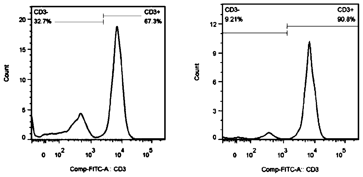
[0028] Enrichment of T lymphocytes (CD3+T) cells in peripheral blood mononuclear cells, including the following steps:

[0029] a. Peripheral blood mononuclear cells (PBMCs) in 50 ml of whole blood were separated by density gradient centrifugation, and the cell density was adjusted to 1×10⁷ cells / ml with phosphate buffered saline (PBS) to make a single cell suspension of PBMCs;

[0030] b. Add FP001 immobilized with CD3 monoclonal antibody (anti-CD3mAb) (a modified polysaccharide, purchased from Nissan Chemical Industries, LTD, item number: 385-07981) and PBS solution into a 50ml cone at a ratio of 1:5. In a centrifuge tube, gently invert and mix evenly to prepare 20ml of solidified anti-CD3mAb three-dimensional antibody network solution;

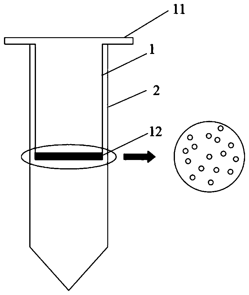
[0031] c. Add the three-dimensional antibody network solution into the inner tube of the cell separation device, and at the same time add the separated PBMCs into the inner tube, gently invert and mix evenly, and incubate at room temperatur...

Embodiment 2

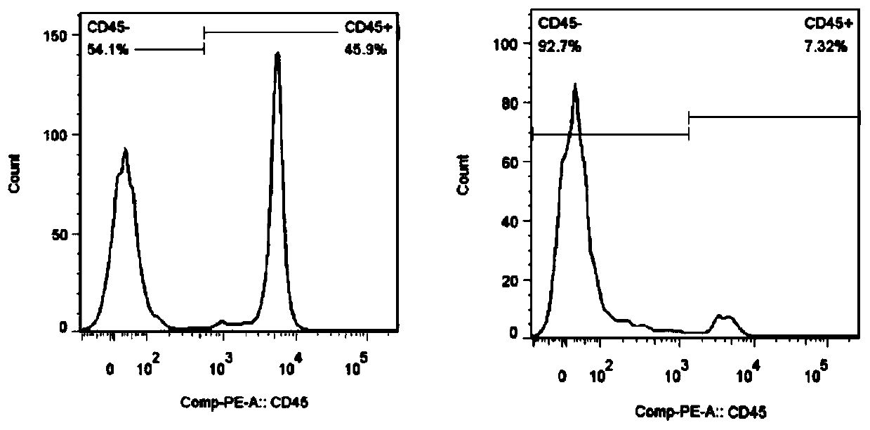
[0036] Depletion of CD45+ cells from peripheral blood mononuclear cells, including the following steps:

[0037] a. Peripheral blood mononuclear cells in 30ml of whole blood were separated by density gradient centrifugation, and the cell density was adjusted to 1×10⁷ cells / ml with PBS solution;

[0038] b. Add FP001 and PBS solution immobilized with CD45 monoclonal antibody into a 50ml conical centrifuge tube at a ratio of 1:6, gently invert and mix evenly to prepare 15ml anti-CD45mAb three-dimensional antibody network solution;

[0039] c. Add the three-dimensional antibody network solution into the inner tube of the cell separation device, and at the same time add the separated PBMCs into the inner tube, gently invert and mix evenly, and incubate at room temperature for 20 minutes;

[0040] d. Centrifuge at 500g for 10 minutes, collect the cell pellet in the outer tube, which is the PBMCs after removing CD45+ cells;

[0041] e, flow cytometry analysis of CD45+ cell removal ...

Embodiment 3

[0042] Enrichment of circulating tumor cells in whole blood, comprising the following steps:

[0043] a. Draw 10ml of peripheral blood from tumor patients for anticoagulation with heparin;

[0044] b. Add the FP001 and PBS solution of solidified epithelial cell adhesion molecule monoclonal antibody (anti-EpCAM mAb) into a 50ml conical centrifuge tube at a ratio of 1:4, gently invert and mix well to prepare 30ml of solidified anti- EpCAM mAb three-dimensional antibody network solution;

[0045] c. Add the three-dimensional antibody network solution into the inner tube of the cell separation device, and at the same time add the separated PBMCs into the inner tube, gently invert and mix evenly, and incubate at room temperature for 20 minutes;

[0046] d. Centrifuge at a centrifugal force of 500g for 10 minutes, add 5ml of PBS to the inner tube to transfer the three-dimensional antibody network and cells trapped by the screen to another conical centrifuge tube;

[0047] e. Add m...


PUM


Abstract

The invention discloses a device for separating cells. The device comprises an inner tube and an outer tube. A flange which extends outwardly is arranged at an opening end of the inner tube, so that the inner tube can be nested in an opening end of the outer tube; a bottom of the inner tube is separated from a bottom of the outer tube by a preset distance, so that single cells which enter the outer tube and are not captured by three-dimensional antibody nets can be accommodated; the bottom of the inner tube is of a porous structure, and the porous structure is used for separating the single cells which are not captured by the three-dimensional antibody nets from the cells which are captured by the three-dimensional antibody nets; the single cells which are not captured by the three-dimensional antibody nets can enter the outer tube via the porous structure, and three-dimensional antibody nets and the cells which are captured by the three-dimensional antibody nets are entrapped in the inner tube by the porous structure. The device for separating the cells has the advantage that specific types of cells can be captured by the three-dimensional antibody nets, and accordingly the cells can be effectively separated from one another.

Description

technical field [0001] The invention relates to the fields of clinical medicine and biological science, in particular to a cell separation device and a cell separation method. Background technique [0002] Cell sorting is the most important technical method and necessary prerequisite for cell biology and molecular biology. At present, the technology is relatively mature and widely used, mainly including: density gradient centrifugation, immune density centrifugation, immunomagnetic bead method and flow cytometry Wait. Among them, density gradient centrifugation is based on the density of cells to achieve the separation of specific types of cells. It is simple to operate and low in cost, but it can only roughly separate cell populations and cannot meet the complex cell separation needs in clinical and scientific research; Density centrifugation, immunomagnetic bead method, and flow sorting are all based on the principle of antibody-captured cells, combined with other technol...

Claims


Application Information

Patent Timeline
IPC IPC(8): C12M1/24C12M1/12C12M1/00C12N5/0783C12N5/09
CPCC12M23/06C12M33/14C12M47/04C12N5/0081C12N5/0636C12N5/0693C12N2509/10
Inventor 姜维吴庆军
Owner SHENZHEN DAKEWE BIOENG
Features
  • Generate Ideas
  • Intellectual Property
  • Life Sciences
  • Materials
  • Tech Scout
  • Unparalleled Data Quality
  • Higher Quality Content
  • 60% Fewer Hallucinations
Learn More