Preparation method for N-doped hollow carbon nanosphere loaded ultra-small gold nanoparticle material

A nanoparticle and hollow carbon technology, applied in the field of biomedical materials, can solve problems such as secondary cancer, large side effects, and immune system damage, and achieve the effects of good biocompatibility, mild reaction conditions, and simple synthesis methods

Inactive Publication Date: 2017-06-13
YANGZHOU UNIV
View PDF1 Cites 21 Cited by
  • Summary
  • Abstract
  • Description
  • Claims
  • Application Information

AI Technical Summary

Problems solved by technology

For example, while killing cancer cells, it will also kill normal cells, causing varying degrees of damage to the human immune system, relatively large side effects, and may even cause secondary cancer in the body

Method used


Image

  • Preparation method for N-doped hollow carbon nanosphere loaded ultra-small gold nanoparticle material
  • Preparation method for N-doped hollow carbon nanosphere loaded ultra-small gold nanoparticle material
  • Preparation method for N-doped hollow carbon nanosphere loaded ultra-small gold nanoparticle material

Examples


Embodiment 1

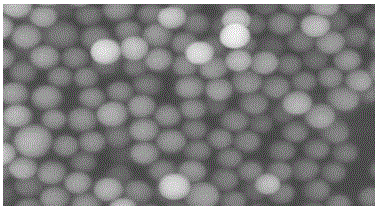
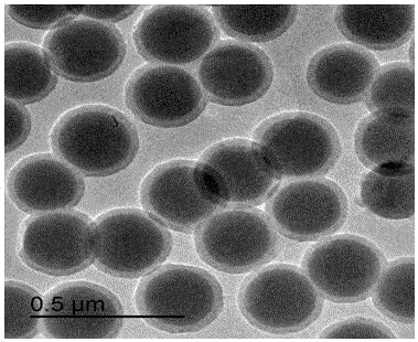
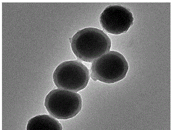
[0035] (1) SiO 2 Preparation of spheres: Measure 60 mL of ethanol, 22.5 mL of deionized water, 1.5 mL of ammonia water and 4.5 mL of LTEOS respectively, and stir at room temperature at 700 r / min for 4 h. Then it was centrifuged at 9000 r / min for 6 min, the solid phase was washed with water and alcohol three times each, and dried at 60 °C to obtain SiO 2 ball.

[0036] (2) SiO 2Preparation of @PDA ball: Weigh SiO 2 Add 100 mg of spheres to 100 mL of Tris aqueous solution (pH=8.5, 0.01 M), sonicate for 10 min at a power of 80 W, then add 100 mg of dopamine hydrochloride, and stir at room temperature at 700 r / min for 10 h. After the reaction was completed, it was centrifuged at 9000 r / min for 6 min, the solid phase was washed with water and alcohol three times each, and dried at 60 °C to obtain SiO 2 @PDA ball.

[0037] (3) SiO 2 Preparation of @N-CNs spheres: SiO 2 @PDA balls were roasted in a tube furnace, and N 2 After exhausting the air in the quartz tube, the temper...

Embodiment 2

[0041] (1) SiO 2 Preparation of spheres: Measure 60 mL of ethanol, 22.5 mL of deionized water, 1.5 mL of ammonia water and 4.5 mL of LTEOS respectively, and stir at 800 r / min for 5 h at room temperature. Then it was centrifuged at 9000 r / min for 6 min, the solid phase was washed with water and alcohol three times each, and dried at 60 °C to obtain SiO 2 ball.

[0042] (2) SiO 2 Preparation of @PDA ball: Weigh SiO 2 Add 100 mg of spheres to 100 mL of Tris aqueous solution (pH=8.5, 0.01 M), sonicate for 8 min at a power of 80 W, then add 200 mg of dopamine hydrochloride, and stir the reaction at room temperature at 800 r / min9 h. After the reaction, centrifuge at 9000 r / min for 6 min, wash the solid phase with water and alcohol three times each, and dry at 60 °C to obtain SiO 2 @PDA ball.

[0043] (3) SiO 2 Preparation of @N-CNs spheres: SiO 2 @PDA balls were roasted in a tube furnace, and N 2 After exhausting the air in the quartz tube, the temperature was raised to 800...

Embodiment 3

[0047] (1) SiO 2 Preparation of spheres: Measure 60 mL of ethanol, 22.5 mL of deionized water, 1.5 mL of ammonia water and 4.5 mL of LTEOS respectively, and stir at room temperature at 600 r / min for 6 h. Then it was centrifuged at 9000 r / min for 6 min, the solid phase was washed with water and alcohol three times each, and dried at 60 °C to obtain SiO 2 ball.

[0048] (2) SiO 2 Preparation of @PDA ball: Weigh SiO 2 Add 100 mg of spheres to 100 mL of Tris aqueous solution (pH=8.5, 0.01 M), sonicate for 5 min at a power of 80 W, then add 100 mg of dopamine hydrochloride, and stir the reaction at room temperature at 600 r / min8 h. After the reaction was completed, it was centrifuged at 9000 r / min for 6 min, the solid phase was washed with water and alcohol three times each, and dried at 60 °C to obtain SiO 2 @PDA ball.

[0049] (3) SiO 2 Preparation of @N-CNs spheres: SiO 2 @PDA balls were roasted in a tube furnace, and N 2 After exhausting the air in the quartz tube, the...


PUM

PropertyMeasurementUnit
particle diameteraaaaaaaaaa

Abstract

The invention relates to a preparation method for an N-doped hollow carbon nanosphere loaded ultra-small gold nanoparticle material and belongs to the field of biomedical materials. The method comprises the following steps: firstly, preparing an SiO2 sphere according to an improved stober method; forming an SiO2@PDA sphere by auto-polymerizing dopamine under a weak basic condition by taking the SiO2 sphere as a template; calcining and charring at high temperature, thereby acquiring an SiO2@N-CNs sphere; preparing SiO2@N-CNs@AuNPs according to the method for reducing HAuCl4 with NaBH4; and finally, performing NaOH etching, thereby acquiring the N-doped hollow carbon nanosphere loaded ultra-small gold nanoparticle material. The synthetic method according to the invention is simple; the reaction conditions are mild; the method is green and environment-friendly; and the prepared material is high in photo-thermal conversion efficiency and photo-thermal stability, good in biocompatibility, low in cost and higher in near infrared absorption capacity.

Description

technical field [0001] The invention belongs to the field of biomedical materials, and in particular relates to photothermal therapy minimally invasive tumor treatment technology. Background technique [0002] The clinical treatment of tumors (cancer) and other diseases has been widely concerned by researchers. In China and some developed countries, the proportion of cancer deaths is as high as 20%, ranking first. Although researchers have done a lot of research work on cancer, the survival rate of cancer patients has only increased by 15%. Today's clinical methods for treating cancer mainly include surgery, radiation therapy, and chemotherapy, all of which have common shortcomings. For example, while killing cancer cells, it will also kill normal cells, causing varying degrees of damage to the immune system of the human body, relatively large side effects, and may even cause secondary cancer in the body. Therefore, how to improve the cure efficiency of cancer patients an...

Claims


Application Information

Patent Timeline
Patent Type & Authority Applications(China)
IPC IPC(8): A61K41/00A61K9/50A61K47/34A61K47/04A61P35/00B82Y5/00
CPCA61K41/0052A61K9/501A61K9/5031A61K9/5073B82Y5/00
Inventor 范磊徐祥东杨莉徐志龙郭荣
Owner YANGZHOU UNIV
Why Eureka
Try Eureka