

Automatic measuring method and device for volume change of skeletal muscle
A volume change and automatic measurement technology, applied in the field of measurement of skeletal muscle volume change, can solve the problems of limited clinical application, radiation, high dose, etc., achieve a wide range of clinical applications, improve processing time, and improve robustness.
- Summary
- Abstract
- Description
- Claims
- Application Information
AI Technical Summary
Problems solved by technology
Method used
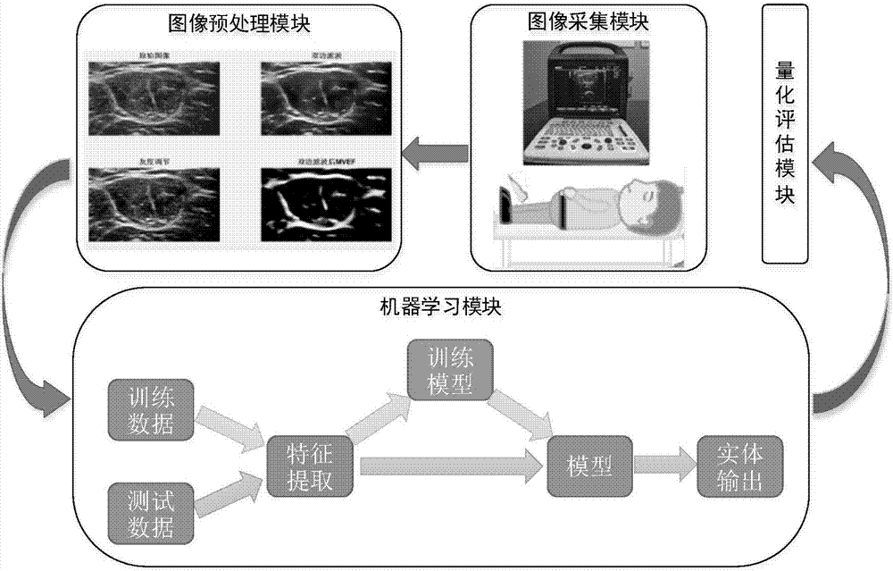
Image

Examples
Embodiment 1
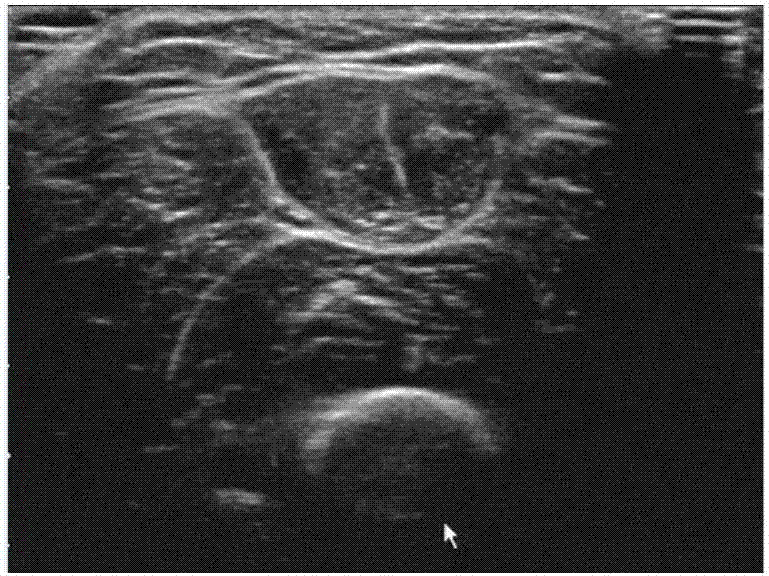
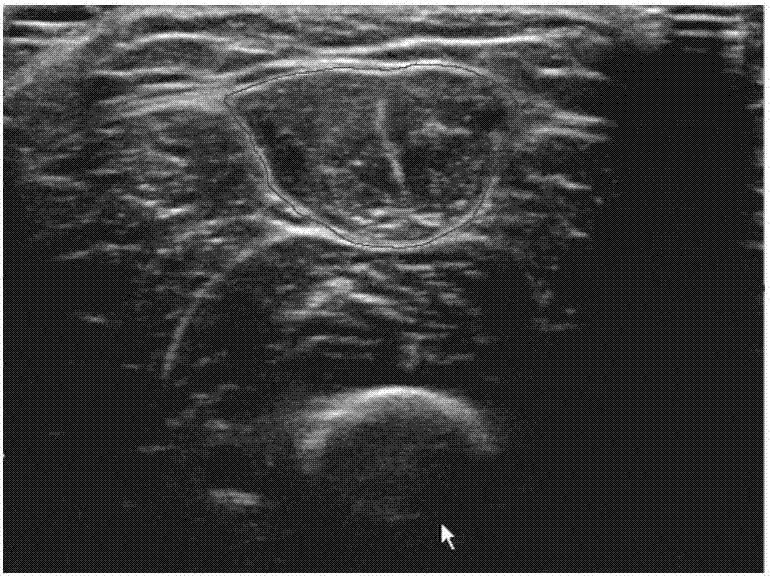
[0047] Take quadriceps atrophy as an example:
[0048] The quadriceps is the largest muscle in the human body and plays a vital role in the daily activities of the human body. In addition, in the statistics of scientific research literature on the combination of ultrasound images with the main lower limb muscles, as shown in Table 1, ultrasound combined with the quadriceps femoris Muscle's literature has the highest citation frequency. Lesions of the thigh muscles, stiffness of the knee joint, and dysfunction of the nerves that control the muscles can all cause a decrease in the activity of the knee joint, thereby reducing the exercise intensity of the quadriceps, resulting in atrophy of the quadriceps and a decrease in muscle strength. A large number of research data show that quadriceps atrophy is more common in patients with knee osteoarthritis, knee cruciate ligament and meniscus injury, etc. Even in healthy people, absolute bed rest can also cause quadriceps muscle weakne...
PUM
 Abstract
Description
Claims
Application Information