Microfluidic-technology-based CTC protein typing kit

A microfluidic technology, protein technology, applied in the field of medicine and biology, CTC protein typing kits, can solve the problems of affecting typing identification, damage cell activity, low recovery rate, etc., to achieve rapid automatic detection and typing, The effect of reducing steric hindrance and improving recycling efficiency

Active Publication Date: 2017-08-01
SUREXAM BIO TECH
View PDF7 Cites 4 Cited by
  • Summary
  • Abstract
  • Description
  • Claims
  • Application Information

AI Technical Summary

Problems solved by technology

Traditional separation methods have defects such as low recovery rate, damaged cell viability, integrity and surface antigen, which affect the subsequent typing and identification

Method used


Image

  • Microfluidic-technology-based CTC protein typing kit
  • Microfluidic-technology-based CTC protein typing kit
  • Microfluidic-technology-based CTC protein typing kit

Examples


Embodiment 1

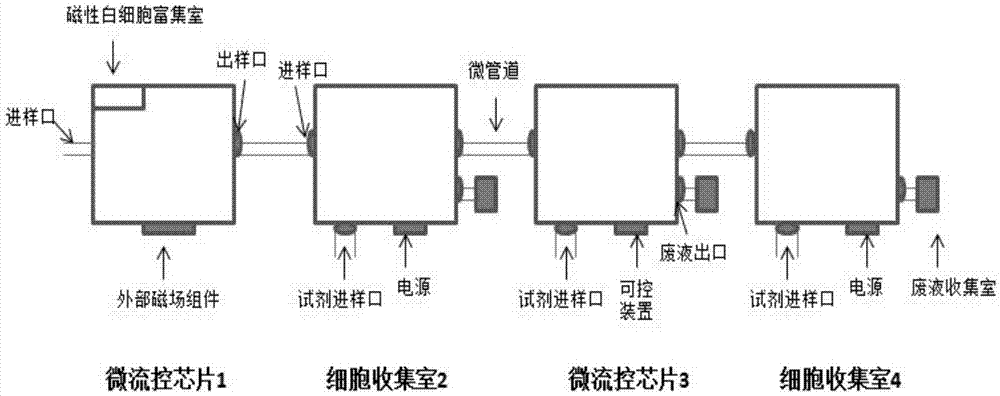
[0032] The CTC protein typing kit based on microfluidic technology described in this embodiment mainly includes:

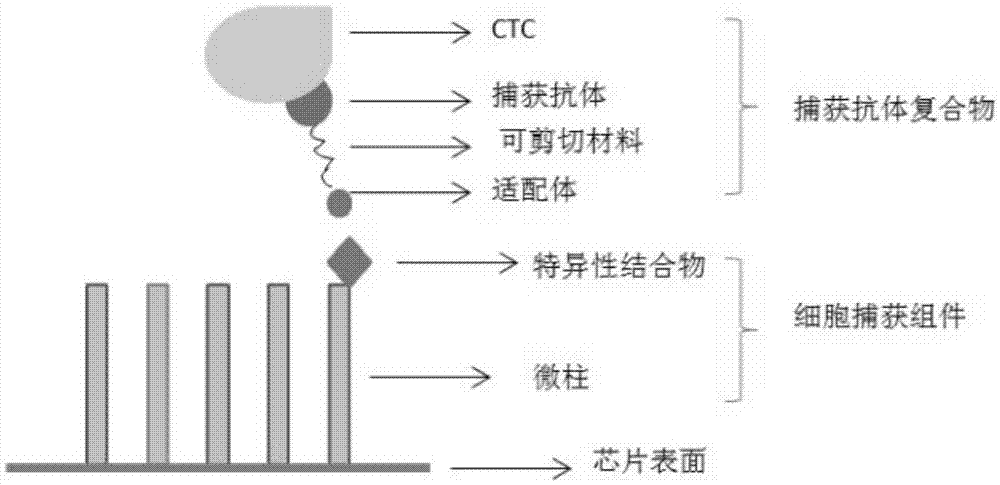
[0033] One, capture antibody complex

[0034] The capture antibody complex includes a capture antibody and an aptamer that specifically bind to each marker protein of CTCs. The capture antibody is used to connect the marker protein and the marker antibody, and can specifically bind to the marker protein and the marker antibody. The capture antibody In order to have high-purity monoclonal antibodies, the reaction characteristics of the selected capture antibodies do not interfere with each other. The capture antibody and the aptamer are connected by a shearable material. The shearable materials include photosensitive materials, temperature-sensitive materials, and humidity-sensitive materials. Materials, heat-sensitive materials, enzymatic degradation materials, etc., aptamers are small molecular compounds that can react with specific binding substances on the cell capt...

Embodiment 2

[0059] Example 2 Using the kit in Example 1 to detect circulating tumor cells in human peripheral blood

[0060] The formulations of the various solutions are as follows:

[0061]

[0062] The capture mixture and the color-developing mixture in this example both use all the antibodies in the corresponding marker protein list in Example 1.

[0063] 1. Sample pretreatment

[0064] 1. Use the preservation solution to store the blood sample in the sample preservation tube, centrifuge at 600×g for 5 min, discard the supernatant, remove the red blood cells, and add 1mL PBS to resuspend.

[0065] 2. Take 450uL of the corresponding magnetic beads and mix, centrifuge to remove the supernatant, add 1mLPBS to resuspend, use 1mg magnetic beads per 30ug magnetic bead antibody, add the magnetic bead antibody mixture to the cell suspension, and react on the mixer for 30min. Centrifuge to remove the supernatant, add 10uLPBS to resuspend.

[0066] 2. Microfluidic device detection

[0067] 1. Add the cell...

Embodiment 3

[0095] Example 3: Using the kit in Example 1 to detect tumor cell lines

[0096] 1. Selection of tumor cell lines

[0097] In this example, we added a known number of cells from the epithelial cell line MCF-10A, the mesenchymal tumor cell line U118, and the epithelial-mesenchymal mixed lung cancer cell line PC-9 to the peripheral blood samples of healthy volunteers, which used The lymphoblast cell line CCRF-HSB-2 was used as a negative control. Those skilled in the art can obtain it through purchase as long as they know the name of the cell line. The kit in Example 1 was used to test the tumor cell capture efficiency of the present invention.

[0098] 2. Sample detection

[0099] Take each of 200 MCF-10A cells, U118 cells and PC-9 cells (determined by a cell counter), add them to 5 mL of healthy volunteers' peripheral blood samples, mix them, and test the samples according to the detection process and method described in Example 2. , The target cell capture rate is calculated by th...


PUM

PropertyMeasurementUnit
Diameteraaaaaaaaaa

Abstract

The invention relates to a microfluidic-technology-based CTC protein typing kit. The kit comprises: (1) capture antibody compounds aiming at different CTCs marker protein, each capture antibody compound being made from a capture antibody in specific binding to corresponding marker protein and an aptamer, wherein the capture antibody and aptamer are connected through a cuttable material which is deoxyribonucleotide double strands having a palindrome structure and two or more than two restriction enzyme sites; and (2) a microfluidic chip, the surface of which is coupled to a cell capture assembly. The cell capture assembly made from a microcolumn is fixed at the surface of the microfluidic chip, and a specific conjugate capable of bonding to the aptamer modifies one end of the microcolumn. Through the microfluidic technology, efficient and fast automatic detection and typing of CTCs is achieved, the kit is simple to operate, false-negative and false-positive results due to man-made operational factors are reduced, and the sensitivity and accuracy of detection are improved.

Description

Technical field [0001] The invention belongs to the field of molecular biology, and relates to medicine and biotechnology, and specifically relates to a CTC protein typing kit based on microfluidic technology, which is suitable for capturing, separating, purifying, and typing identification of circulating tumor cells in blood. Background technique [0002] Circulating tumor cells (CTCs) originate from primary tumors or metastatic tumors, gain the ability to break away from the basement membrane and enter blood vessels through invading tissue matrix. It is a collective term for various types of tumor cells that exist in the peripheral blood. According to the expression difference of its surface antigens, it is mainly divided into epithelial marker positive phenotype (referred to as epithelial cell type), mesenchymal marker positive phenotype (referred to as mesenchymal cell type), and epithelial and mesenchymal mixed phenotype (referred to as epithelial-interstitial cell type). Pl...

Claims


Application Information

Patent Timeline
IPC IPC(8): G01N33/68G01N33/574
CPCG01N33/5304G01N33/5308G01N33/56966G01N33/574G01N33/6854
Inventor 刘志明吴诗扬廖传荣
Owner SUREXAM BIO TECH
Why Eureka
Try Eureka