
Container for vacuum collection of karyotype analysis sample

A chromosome karyotype and collection container technology, applied in the field of vacuum collection containers, can solve problems such as being susceptible to contamination, and achieve the effect of eliminating intermediate links and improving efficiency

Inactive Publication Date: 2017-08-08
付士明
View PDF6 Cites 1 Cited by
  • Summary
  • Abstract
  • Description
  • Claims
  • Application Information

AI Technical Summary

Problems solved by technology

[0003] The purpose of the present invention is to overcome the defect that the sample collection operation of the current karyotype analysis is complicated and easy to be polluted, and to invent a simple and easy vacuum collection container for karyotype analysis samples that can be directly used in subsequent experiments after sample collection

Method used


Image

  • Container for vacuum collection of karyotype analysis sample
  • Container for vacuum collection of karyotype analysis sample
  • Container for vacuum collection of karyotype analysis sample

Examples


Embodiment approach

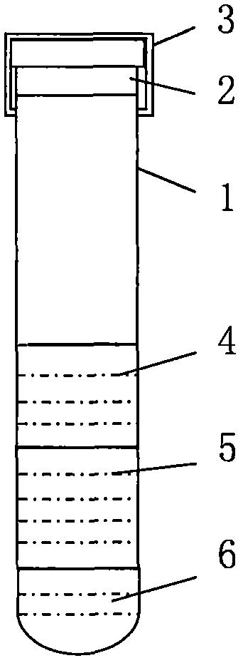
[0015] 1. The original tube 1 is provided with a cell culture medium 4, a blood anticoagulant 5, and a mitogen 6, and the cell culture medium 4, the blood anticoagulant 5, and the mitogen 6 are mixed as a medium mixture I.

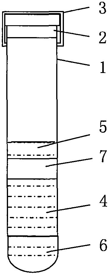
[0016] 2. The original tube 1 is provided with a blood anticoagulant 5, a blood separation gel 7, a cell culture medium 4 and a mitogen 6, wherein the cell culture medium 4 and the mitogen 6 are mixed as the medium mixture II.

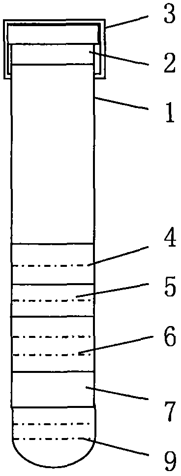
[0017] 3. The original tube 1 is provided with cell culture medium 4, blood anticoagulant 5, mitogen 6, blood separation gel 7 and leukocyte separation liquid 9, wherein cell culture medium 4, blood anticoagulant 5 and mitogen 6 After mixing, it was used as medium mixture III.

[0018] Four. The original tube 1 is provided with cell culture medium 4, blood anticoagulant 5, mitogen 6, blood separation gel 7 and mitosis inhibitor 8, wherein cell culture medium 4, blood anticoagulant 5 and mitogen 6 mixed as medium mixture IV.

[00...

Embodiment 1

[0020] The method of use in Example 1: directly collect the peripheral blood of the test subject and then directly culture the whole blood. After the culture is completed, mitosis inhibitors are added for subsequent experiments.

Embodiment 2

[0021] The usage method of embodiment two: directly collect the experimenter's peripheral blood, then centrifuge through centrifuge, because the specific gravity of blood separation gel is smaller than red blood cells but greater than white blood cells and culture medium mixture and plasma, so the red blood cells are pressed under the separation gel, the separation gel The above is a mixture of leukocytes, medium mixture, mitogen and plasma. After centrifugation, leukocytes are cultured. After the culture is completed, mitosis inhibitors are added for subsequent experiments.


PUM


Abstract

The invention discloses a container for vacuum collection of a karyotype analysis sample. The container comprises a primitive tube, a rubber plug and a protective cap, wherein the primitive tube, the rubber plug and the protective cap are assembled into the vacuated collection container. The container is characterized in that a cell medium, a blood anticoagulant, a mitogen, a serum separating medium, a mitosis repressor and a leukocyte separating medium are arranged in the primitive tube; the cell medium comprises RPMI-1640 or 199 or F10 or Eagle; the blood anticoagulant is heparinate, sodium citrate and buffer solution thereof, EDTA salt, blood preservation solution and hirudin; the mitogen is phytolectin (PHA) or concanavalin-A; and the mitosis repressor is demecolcine, colchicine and derivatives thereof. The collection container not only can be used for karyotype analysis, but also can be used for sample collection for in situ chromosomal hybridization, chromosome linkage map analysis and microseparation and cloning. The container is simple and easy to operate, and samples can be directly used for follow-up experiments after being collected.

Description

technical field [0001] The invention relates to a vacuum collection container, in particular to a vacuum collection container for chromosome karyotype analysis samples. Background technique [0002] Chromosomal karyotype analysis is the basic method of cytogenetics research, and it is one of the indispensable and important means to study the relationship between species evolution, classification and chromosome structure, shape and function. Chromosomal karyotype analysis is achieved through peripheral blood samples, amniocentesis samples, or tissue homogenate samples. Therefore, for chromosomal karyotype analysis, samples must first be collected. Since the collected samples cannot be directly used for follow-up experiments, it is also necessary to Subsequent processing of the sample includes culturing the sample, performing colchicine treatment and hypotonic treatment on the sample, fixing the sample with a fixative, etc. The entire operation process is very complicated and ...

Claims


Application Information

Patent Timeline
Patent Type & Authority Applications(China)
IPC IPC(8): C12M1/26C12M1/24
CPCB01L3/5021
Inventor 付士明
Owner 付士明
Learn More