RNCEC (rabbit normal corneal epithelial cells) and application thereof

A corneal epithelial cell, normal technology, applied in the field of cell biology, can solve the problems of not being able to be passed down, killing experimental animals, etc.

Inactive Publication Date: 2017-08-18
SHENZHEN EYE HOSPITAL
View PDF2 Cites 1 Cited by
  • Summary
  • Abstract
  • Description
  • Claims
  • Application Information

AI Technical Summary

Problems solved by technology

At present, there is no rabbit corneal epithelial cell model used in in vitro drug susceptibility and toxicity testing in the world, and the existing research is only carried out on the b

Method used


Image

  • RNCEC (rabbit normal corneal epithelial cells) and application thereof
  • RNCEC (rabbit normal corneal epithelial cells) and application thereof
  • RNCEC (rabbit normal corneal epithelial cells) and application thereof

Examples


Embodiment 1

[0049] [Example 1] Primary isolation and culture method of rabbit normal corneal epithelial cells

[0050] 1) Strictly follow the animal experiment procedures and regulations of ARRIVE (Animal Research: Reporting of In Vivo Experiments), and collect normal rabbit corneal samples;

[0051] 2) Preparation of digestion solution: HL medium containing 0.2 mg / mL of collagenase and dispase; among them, HL medium is: DMEM (GIBCO # 11965-092) and serum-free medium SFM (GIBCO # 10744-019 ) were mixed at a volume ratio of 1 : 3, and 5% (v / v) fetal bovine serum was added, as well as 0.4 μg / mL cortisol (hydrocortisone), 5 μg / mL insulin (insulin), and 8.4 ng / mL cholera toxin ( cholera toxin), 10 ng / mL epithelial growth factor (EGF), 24 μg / mL adenine, 100 U / mL penicillin, 100 μg / mL streptomycin, 0.25 μg / mL amphotericin B (Fungizone), 30 μM Fasudil (Fasudil), the above medium needs to be filtered through a 0.22 μm pore size filter).

[0052] 3) Wash the isolated fresh tissue once with 95-10...

Embodiment 2

[0061] [Example 2] Subculture method of rabbit normal corneal epithelial cells RNCEC / HL-032

[0062] 1) When the rabbit normal corneal epithelial cells proliferate to 70-90% abundance, wash the cells twice with 1xPBS, and digest the monolayer cells with 0.05% trypsin / EDTA for 2-5 minutes.

[0063] 2) 10ml of complete DMEM to neutralize the digestion reaction.

[0064] 3) Centrifuge at 1000rmp for 5 minutes to remove the supernatant.

[0065] 4) At a ratio of 1:2, as long as the cells can adhere to the wall and grow normally), resuspend the cell pellet in 10ml of the medium in Example 1 for inoculation.

[0066] 5) If necessary, the 1x10 6 Corneal epithelial cells were resuspended in 1-2ml of cell freezing medium (90% fetal bovine serum and 10% DMSO), and stored in liquid nitrogen for future use.

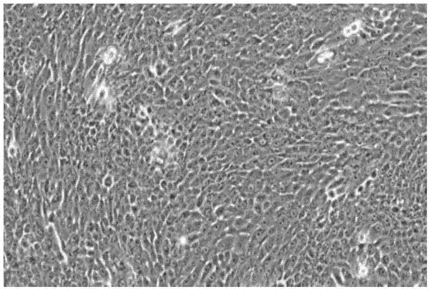
[0067] Rabbit normal corneal epithelial cells subcultured according to the above method, the growth curve is as follows: figure 2 , the cells maintain a steady state of prolifer...

Embodiment 3

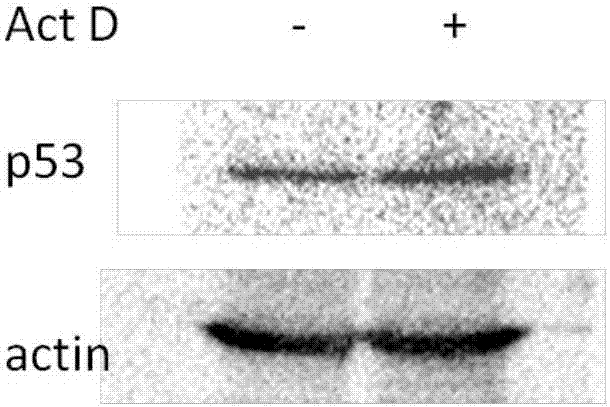
[0068] [Example 3] DNA damage response experiment of rabbit normal corneal epithelial cells RNCEC / HL-032

[0069] 1) Normal rabbit corneal epithelial cells were digested with 0.05% trypsin, prepared into a single cell suspension, inoculated into a 6-well plate, 1x10 per well 6 cells at 37°C, 5% CO 2 Incubator cultivation.

[0070] 2) Add 10 μM actinomycin D (Act D), at 37°C, 5% CO 2 Incubator for 24 hours.

[0071] 3) Cells were collected, lysed with 200 μl RIPA lysate on ice for 30 minutes, centrifuged at 12000 rpm at 4°C for 30 minutes, and the supernatant was collected.

[0072] 4) Perform western blot detection on the collected protein samples.

[0073] The result is as image 3 As shown, after the normal rabbit corneal epithelial cells were treated with Act D, the expression of p53 protein was up-regulated, indicating that the cells had a normal response ability to DNA damage.


PUM


Abstract

The invention discloses RNCEC (rabbit normal corneal epithelial cells) and an application thereof. The cell is named as RNCEC/HL-032, and the collection number is CCTCC NO: C201677. A primary isolation culture method of the cell is as follows: a normal corneal limbus tissue sample of a New Zealand rabbit is digested, then dispase and DNase I are added after digestion, cells are filtered and collected in a centrifugal manner and then resuspended with an HL culture medium for inoculated culture. The subculturing method of RNCEC comprises steps as follows: the cells are digested with pancreatin-EDTA when proliferating to have the abundance of 70%-90% and then neutralized with a DMEM (dulbecco's modified eagle medium); the cells are collected in a centrifugal manner and resuspended with the HL culture medium for inoculated culture. A 2D and 3D culturing system established for the cell can be used for animal substitutive experiments of corneal drugs and used for detecting pharmacology and toxicology of drugs and establishing a new experimental animal substitution model for corneal drug research.

Description

technical field [0001] The invention belongs to the field of cell biology, and relates to a rabbit normal corneal epithelial cell and its primary separation culture and subculture method and application. Background technique [0002] Modern medicine is developed on the basis of animal experiments. Whether it is the research and development of vaccines, organ transplantation, research on physiology, pharmacology, toxicology, or the development of cosmetics, all come with the sacrifice of experimental animals. If animal experiments are canceled, the development of modern medicine will stagnate, and it will be impossible for humans to overcome diseases. At present, an internationally recognized principle for animal experiments is "replacement, reduction, and optimization." First of all, researchers should try their best to seek alternatives to animal experiments, for example, using computer dynamic simulation and cell tissue culture plasma technology. Among them, cell tissue...

Claims


Application Information

Patent Timeline
IPC IPC(8): C12N5/071C12N5/079C12Q1/02
CPCC12N5/0625C12N5/0621C12N2513/00G01N33/5014G01N33/5058
Inventor 叶琳李晖王玲魏高斌叶立娜王媛殷国干
Owner SHENZHEN EYE HOSPITAL
Why Eureka
Try Eureka