Method for observing same sample by using paraffin section and scanning electron microscope

A scanning electron microscope and paraffin section technology, applied in the field of biological experiments, can solve the problems of inability to obtain a three-dimensional outline of plant tissue, waste of materials, inability to see the results of paraffin sections, etc., and achieve favorable effects of plant morphological development.

Active Publication Date: 2017-08-29
NORTHWEST A & F UNIV
View PDF4 Cites 2 Cited by
  • Summary
  • Abstract
  • Description
  • Claims
  • Application Information

AI Technical Summary

Problems solved by technology

However, if two samples with similar developmental status are found for processing, then the problem is that it is impossible to have two completely identical samples, and only approximately identical results can be obtained, and there is a large waste of materials
If the wax strip is dewaxed, the disadvantage is that only the scanned image of the cut surface can be obtained, and the results of the paraffin section cannot be seen; at the same time, the three-dimensional outline of the plant tissue cannot be obtained because the sample has been sectioned

Method used


Image

  • Method for observing same sample by using paraffin section and scanning electron microscope
  • Method for observing same sample by using paraffin section and scanning electron microscope
  • Method for observing same sample by using paraffin section and scanning electron microscope

Examples


Embodiment 1

[0026] The making of embodiment 1 paraffin section

[0027] The material is magnolia seeds

[0028] 1) Fixation: use FAA fixative solution (70% alcohol: formaldehyde: glacial acetic acid = 90:5:5), fix at room temperature for 24 hours.

[0029] 2) Soak in dehydration series solution, soak each reagent for one day, follow the order of Table 1-6.

[0030]

[0031]

[0032] 3) Dip wax

[0033] Adjust the temperature of the incubator to 60°C, put the sample in a glass bottle, add an appropriate amount of wax shavings, a small amount of tert-butanol, cover it, open the cover two days later, let the tert-butanol volatilize, and soak in the wax for a week.

[0034] 4) Embedding

[0035] Pour the liquid wax into the aluminum box and place it on the spreading machine to prevent solidification. The temperature of the spreading machine is set at 60°C. Put the material soaked in wax for a week into the liquid wax, and use hot tweezers when slowly solidifying at room temperature ...

Embodiment 2

[0047] Embodiment 2 dewaxing method and scanning electron microscope

[0048] Immersion dewaxing:

[0049] 1. Dewaxing--Reagent: xylene or tert-butanol

[0050] The remaining samples of the paraffin sections were divided into two groups, one group was soaked in xylene and placed in a 40°C incubator; the other group was soaked in tert-butanol and placed in a 60°C incubator, and the soaking time was set to 3 hours, 6 hours and 9 hours, and two samples for each time period were replicated three times.

[0051] 2. Transition to isoamyl acetate

[0052]

[0053] 3. Critical point drying

[0054] Put the material in a critical point dryer for critical point drying

[0055] 4. Electron microscope scanning

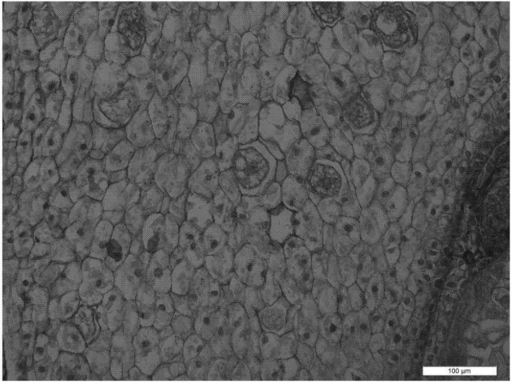
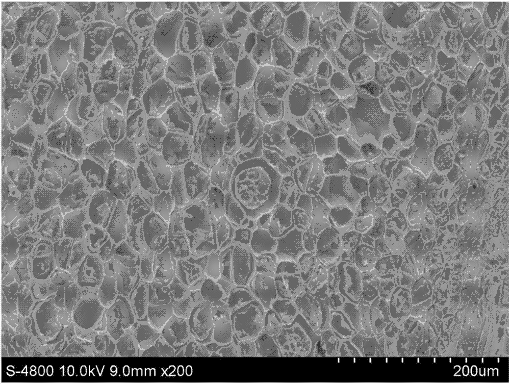
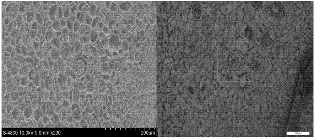
[0056] see attached results Figure 2-3 , Figure 8-14 .


PUM


Abstract

The invention discloses a method for observing a same sample by using a paraffin section and a scanning electron microscope. The method comprises the following steps: subjecting the sample to paraffin embedding and then carrying out tissue sectioning until the symmetric axis of the sample is reached, or according to observation of tissue sectioning results; then subjecting the rest embedded paraffin block of the sample to deparaffinization; and then carrying out scanning via the electron microscope so as to obtain electron microscope scanning results of a sample section. According to the invention, one same sample is made full use; the morphologies of a variety of cells in the sample can be observed, and the outer contour of tissue and high-magnification high-resolution three-dimensional images of tiny structures in the tissue can be observed in the obtained electron microscope scanning results of the sample at the same time, which are favorable for research on the morphological development of plants.

Description

technical field [0001] The invention belongs to the technical field of biological experiments, in particular to a method for observing the same sample by using a paraffin section and a scanning electron microscope. Background technique [0002] The paraffin section and scanning electron microscope observation techniques of plant tissue have become mature, but two observation methods for one sample are not common. [0003] Paraffin section is to cut the biological tissue into thin slices, and then observe the outline of the tissue section and the morphology of various cells in the tissue through optical microscope observation. The obtained image has the characteristics of wide field of view, thick lines, and large outline. Planar image and optical microscope magnification is limited (maximum magnification 1000 times), it is difficult to observe the overall shape, three-dimensional shape and subcellular structure of plant tissues. Scanning electron microscope technology can w...

Claims


Application Information

Patent Timeline
Patent Type & Authority Applications(China)
IPC IPC(8): G01N23/22G01N1/28
CPCG01N23/2202
Inventor 张鑫赵忠江国彬张一帆
Owner NORTHWEST A & F UNIV
Why Eureka
Try Eureka